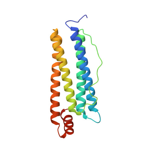
2Z5Q

Apo-Fr with intermediate content of Pd ion


Experimental Data Snapshot

  • Method: X-RAY DIFFRACTION
  • Resolution: 2.10 Å
  • R-Value Free: 0.254 
  • R-Value Work: 0.199 
  • R-Value Observed: 0.202 

Starting Model: experimental
View more details

wwPDB Validation   3D Report Full Report


This is version 1.2 of the entry. See complete history


Literature

Process of accumulation of metal ions on the interior surface of apo-ferritin: crystal structures of a series of apo-ferritins containing variable quantities of Pd(II) ions.

Ueno, T.Abe, M.Hirata, K.Abe, S.Suzuki, M.Shimizu, N.Yamamoto, M.Takata, M.Watanabe, Y.

(2009) J Am Chem Soc 131: 5094-5100

  • DOI: https://doi.org/10.1021/ja806688s
  • Primary Citation of Related Structures:  
    2Z5P, 2Z5Q, 2Z5R, 3FI6

  • PubMed Abstract: 

    Accumulation of metal ions on protein surfaces is an important subject in the field of materials science because these processes are applicable to the preparation of bioinspired inorganic materials. While previous studies related to this subject have focused on the preparation of nanomaterials using protein scaffolds, the detailed processes of metal ion deposition and metal core formation on a protein surface require clarification. Elucidation of the coordination structures of multinuclear metal binding sites on proteins at an early stage as well as intermediate and fully occupied stages of the metal ion deposition will help us to understand the reaction mechanisms so that desirable inorganic materials can be prepared using protein scaffolds. In this Article, we report on the detailed processes of accumulation of Pd(II) ions demonstrated by a series of X-ray crystal structural analyses of apo-ferritin (apo-Fr), an iron storage protein, containing different amounts of Pd(II) ions in the protein cage. We have identified the specific binding sites of Pd(II) ions and analyzed the dynamic changes in the coordination structure by a combination of the crystal structures and ICP quantitative analyses of apo-Fr containing low, intermediate, and high content of Pd(II) ions. Our studies on Pd(II).apo-Frs provide intriguing implications for the preparation of many other inorganic materials using protein surfaces.


  • Organizational Affiliation

    Institute for Integrated Cell-Material Sciences, Funai Center, Kyoto University, Katsura, Nishikyo-ku, Kyoto 615-8510, Japan. taka@sbchem.kyoto-u.ac.jp


Macromolecules
Find similar proteins by:  (by identity cutoff)  |  3D Structure
Entity ID: 1
MoleculeChains Sequence LengthOrganismDetailsImage
Ferritin light chain174Equus caballusMutation(s): 0 
Gene Names: FTL
UniProt
Find proteins for P02791 (Equus caballus)
Explore P02791 
Go to UniProtKB:  P02791
Entity Groups  
Sequence Clusters30% Identity50% Identity70% Identity90% Identity95% Identity100% Identity
UniProt GroupP02791
Sequence Annotations
Expand
  • Reference Sequence
Small Molecules
Ligands 3 Unique
IDChains Name / Formula / InChI Key2D Diagram3D Interactions
CD
Query on CD

Download Ideal Coordinates CCD File 
K [auth A]CADMIUM ION
Cd
WLZRMCYVCSSEQC-UHFFFAOYSA-N
PD
Query on PD

Download Ideal Coordinates CCD File 
B [auth A]
C [auth A]
D [auth A]
E [auth A]
F [auth A]
B [auth A],
C [auth A],
D [auth A],
E [auth A],
F [auth A],
G [auth A],
H [auth A],
I [auth A],
J [auth A]
PALLADIUM ION
Pd
MUJIDPITZJWBSW-UHFFFAOYSA-N
GOL
Query on GOL

Download Ideal Coordinates CCD File 
L [auth A],
M [auth A]
GLYCEROL
C3 H8 O3
PEDCQBHIVMGVHV-UHFFFAOYSA-N
Experimental Data & Validation

Experimental Data

  • Method: X-RAY DIFFRACTION
  • Resolution: 2.10 Å
  • R-Value Free: 0.254 
  • R-Value Work: 0.199 
  • R-Value Observed: 0.202 
  • Space Group: F 4 3 2
Unit Cell:
Length ( Å )Angle ( ˚ )
a = 180.57α = 90
b = 180.57β = 90
c = 180.57γ = 90
Software Package:
Software NamePurpose
REFMACrefinement
HKL-2000data collection
SCALEPACKdata scaling
MOLREPphasing

Structure Validation

View Full Validation Report


Entry History 

Deposition Data

Revision History  (Full details and data files)

  • Version 1.0: 2008-07-29
    Type: Initial release
  • Version 1.1: 2011-07-13
    Changes: Non-polymer description, Version format compliance
  • Version 1.2: 2023-11-01
    Changes: Data collection, Database references, Derived calculations, Refinement description