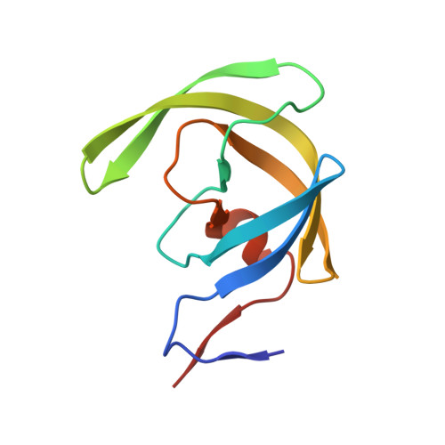
3BVA

Cystal structure of HIV-1 Active Site Mutant D25N and p2-NC analog inhibitor


Experimental Data Snapshot

  • Method: X-RAY DIFFRACTION
  • Resolution: 1.05 Å
  • R-Value Free: 0.180 
  • R-Value Work: 0.150 
  • R-Value Observed: 0.155 

Starting Model: experimental
View more details

wwPDB Validation   3D Report Full Report


Ligand Structure Quality Assessment 


This is version 1.4 of the entry. See complete history


Literature

Effect of the Active Site D25N Mutation on the Structure, Stability, and Ligand Binding of the Mature HIV-1 Protease.

Sayer, J.M.Liu, F.Ishima, R.Weber, I.T.Louis, J.M.

(2008) J Biol Chem 283: 13459-13470

  • DOI: https://doi.org/10.1074/jbc.M708506200
  • Primary Citation of Related Structures:  
    3BVA, 3BVB

  • PubMed Abstract: 

    All aspartic proteases, including retroviral proteases, share the triplet DTG critical for the active site geometry and catalytic function. These residues interact closely in the active, dimeric structure of HIV-1 protease (PR). We have systematically assessed the effect of the D25N mutation on the structure and stability of the mature PR monomer and dimer. The D25N mutation (PR(D25N)) increases the equilibrium dimer dissociation constant by a factor >100-fold (1.3 +/- 0.09 microm) relative to PR. In the absence of inhibitor, NMR studies reveal clear structural differences between PR and PR(D25N) in the relatively mobile P1 loop (residues 79-83) and flap regions, and differential scanning calorimetric analyses show that the mutation lowers the stabilities of both the monomer and dimer folds by 5 and 7.3 degrees C, respectively. Only minimal differences are observed in high resolution crystal structures of PR(D25N) complexed to darunavir (DRV), a potent clinical inhibitor, or a non-hydrolyzable substrate analogue, Ac-Thr-Ile-Nle-r-Nle-Gln-Arg-NH(2) (RPB), as compared with PR.DRV and PR.RPB complexes. Although complexation with RPB stabilizes both dimers, the effect on their T(m) is smaller for PR(D25N) (6.2 degrees C) than for PR (8.7 degrees C). The T(m) of PR(D25N).DRV increases by only 3 degrees C relative to free PR(D25N), as compared with a 22 degrees C increase for PR.DRV, and the mutation increases the ligand dissociation constant of PR(D25N).DRV by a factor of approximately 10(6) relative to PR.DRV. These results suggest that interactions mediated by the catalytic Asp residues make a major contribution to the tight binding of DRV to PR.


  • Organizational Affiliation

    Laboratory of Chemical Physics, NIDDK, National Institutes of Health, Bethesda, Maryland 20892-0520, USA.


Macromolecules
Find similar proteins by:  (by identity cutoff)  |  3D Structure
Entity ID: 1
MoleculeChains Sequence LengthOrganismDetailsImage
Protease (Retropepsin)
A, B
99Human immunodeficiency virus 1Mutation(s): 6 
Gene Names: GAG-POL
EC: 3.4.23.16
UniProt
Find proteins for P03367 (Human immunodeficiency virus type 1 group M subtype B (isolate BRU/LAI))
Explore P03367 
Go to UniProtKB:  P03367
Entity Groups  
Sequence Clusters30% Identity50% Identity70% Identity90% Identity95% Identity100% Identity
UniProt GroupP03367
Sequence Annotations
Expand
  • Reference Sequence
Small Molecules
Binding Affinity Annotations 
IDSourceBinding Affinity
2NG PDBBind:  3BVA Kd: 300 (nM) from 1 assay(s)
Experimental Data & Validation

Experimental Data

  • Method: X-RAY DIFFRACTION
  • Resolution: 1.05 Å
  • R-Value Free: 0.180 
  • R-Value Work: 0.150 
  • R-Value Observed: 0.155 
  • Space Group: P 21 21 2
Unit Cell:
Length ( Å )Angle ( ˚ )
a = 58.196α = 90
b = 85.893β = 90
c = 46.547γ = 90
Software Package:
Software NamePurpose
SHELXmodel building
SHELXL-97refinement
HKL-2000data collection
HKL-2000data reduction
HKL-2000data scaling
AMoREphasing

Structure Validation

View Full Validation Report


Ligand Structure Quality Assessment 


Entry History 

Deposition Data

Revision History  (Full details and data files)

  • Version 1.0: 2008-04-01
    Type: Initial release
  • Version 1.1: 2011-07-13
    Changes: Atomic model, Database references, Derived calculations, Non-polymer description, Structure summary, Version format compliance
  • Version 1.2: 2013-02-27
    Changes: Other
  • Version 1.3: 2021-10-20
    Changes: Database references, Derived calculations
  • Version 1.4: 2023-08-30
    Changes: Data collection, Refinement description