3CEL

ACTIVE-SITE MUTANT E212Q DETERMINED AT PH 6.0 WITH CELLOBIOSE BOUND IN THE ACTIVE SITE


Experimental Data Snapshot

  • Method: X-RAY DIFFRACTION
  • Resolution: 2.00 Å
  • R-Value Free: 0.256 
  • R-Value Work: 0.200 
  • R-Value Observed: 0.200 

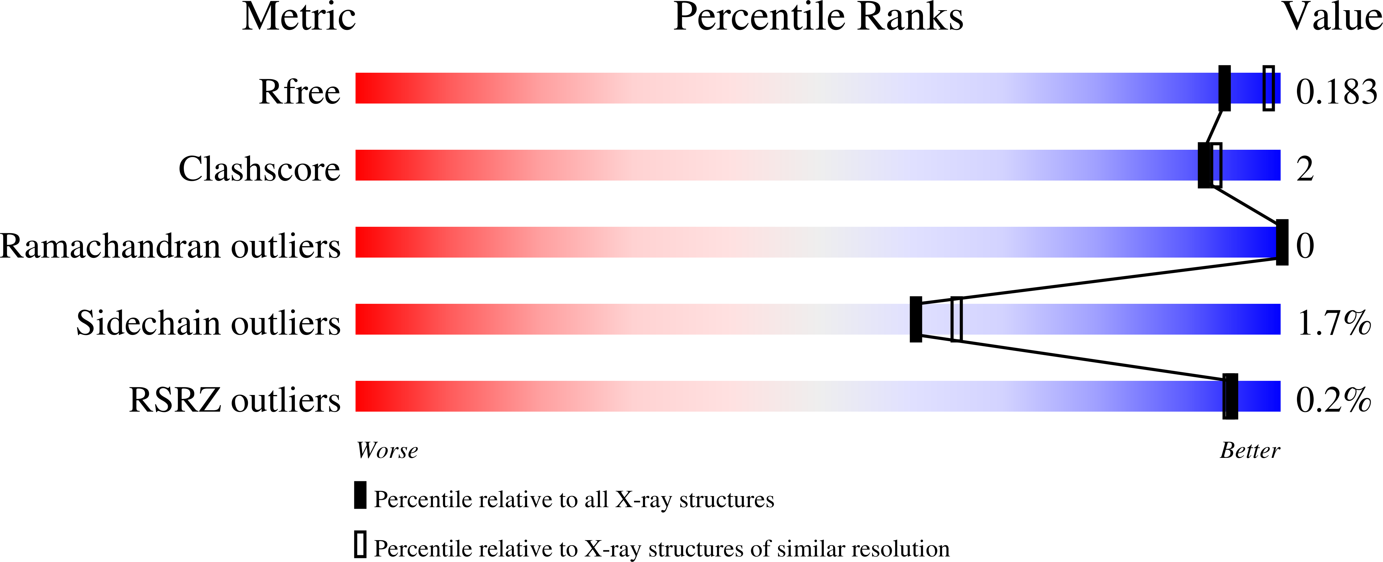
wwPDB Validation   3D Report Full Report


Ligand Structure Quality Assessment 


This is version 3.2 of the entry. See complete history


Literature

Activity studies and crystal structures of catalytically deficient mutants of cellobiohydrolase I from Trichoderma reesei.

Stahlberg, J.Divne, C.Koivula, A.Piens, K.Claeyssens, M.Teeri, T.T.Jones, T.A.

(1996) J Mol Biol 264: 337-349

  • DOI: https://doi.org/10.1006/jmbi.1996.0644
  • Primary Citation of Related Structures:  
    2CEL, 3CEL, 4CEL

  • PubMed Abstract: 

    The roles of the residues in the catalytic trio Glu212-Asp214-Glu217 in cellobiohydrolase I (CBHI) from Trichoderma reesei have been investigated by changing these residues to their isosteric amide counterparts. Three mutants, E212Q, D214N and E217Q, were constructed and expressed in T. reesei. All three point mutations significantly impair the catalytic activity of the enzyme, although all retain some residual activity. On the small chromophoric substrate CNP-Lac, the kcat values were reduced to 1/2000, 1/85 and 1/370 of the wild-type activity, respectively, whereas the KM values remained essentially unchanged. On insoluble crystalline cellulose, BMCC, no significant activity was detected for the E212Q and E217Q mutants, whereas the D214N mutant retained residual activity. The consequences of the individual mutations on the active-site structure were assessed for two of the mutants, E212Q and D214N, by X-ray crystallography at 2.0 A and 2.2 A resolution, respectively. In addition, the structure of E212Q CBHI in complex with the natural product, cellobiose, was determined at 2.0 A resolution. The active-site structure of each mutant is very similar to that of the wild-type enzyme. In the absence of ligand, the active site of the D214N mutant contains a calcium ion firmly bound to Glu212, whereas that of E212Q does not. This supports our hypothesis that Glu212 is the charged species during catalysis. As in the complex of wild-type CBHI with bound o-iodobenzyl-1-thio-beta-D-glucoside, cellobiose is bound to the two product sites in the complex with E212Q. However, the binding of cellobiose differs from that of the glucoside in that the cellobiose is shifted away from the trio of catalytic residues to interact more intimately with a loop that is part of the outer wall of the active site.


  • Organizational Affiliation

    Department of Molecular Biology, University of Uppsala, Sweden.


Macromolecules
Find similar proteins by:  (by identity cutoff)  |  3D Structure
Entity ID: 1
MoleculeChains Sequence LengthOrganismDetailsImage
1,4-BETA-D-GLUCAN CELLOBIOHYDROLASE I434Trichoderma reeseiMutation(s): 1 
Gene Names: CBH1
EC: 3.2.1.91
UniProt
Find proteins for P62694 (Hypocrea jecorina)
Explore P62694 
Go to UniProtKB:  P62694
Entity Groups  
Sequence Clusters30% Identity50% Identity70% Identity90% Identity95% Identity100% Identity
UniProt GroupP62694
Glycosylation
Glycosylation Sites: 1
Sequence Annotations
Expand
  • Reference Sequence
Oligosaccharides

Help

Entity ID: 2
MoleculeChains Length2D Diagram Glycosylation3D Interactions
beta-D-glucopyranose-(1-4)-beta-D-glucopyranose
B
2N/A
Glycosylation Resources
GlyTouCan:  G84824ZO
GlyCosmos:  G84824ZO
Biologically Interesting Molecules (External Reference) 1 Unique
Experimental Data & Validation

Experimental Data

  • Method: X-RAY DIFFRACTION
  • Resolution: 2.00 Å
  • R-Value Free: 0.256 
  • R-Value Work: 0.200 
  • R-Value Observed: 0.200 
  • Space Group: I 2 2 2
Unit Cell:
Length ( Å )Angle ( ˚ )
a = 84α = 90
b = 84.1β = 90
c = 111.5γ = 90
Software Package:
Software NamePurpose
DENZOdata reduction
CCP4data reduction
X-PLORmodel building
X-PLORrefinement
CCP4data scaling
X-PLORphasing

Structure Validation

View Full Validation Report


Ligand Structure Quality Assessment 


Entry History 

Deposition Data

Revision History  (Full details and data files)

  • Version 1.0: 1997-03-12
    Type: Initial release
  • Version 1.1: 2008-03-25
    Changes: Version format compliance
  • Version 1.2: 2011-07-13
    Changes: Non-polymer description, Version format compliance
  • Version 2.0: 2019-12-25
    Changes: Atomic model, Data collection, Database references, Derived calculations, Polymer sequence
  • Version 3.0: 2020-07-29
    Type: Remediation
    Reason: Carbohydrate remediation
    Changes: Atomic model, Data collection, Derived calculations, Structure summary
  • Version 3.1: 2021-11-03
    Changes: Database references, Derived calculations, Structure summary
  • Version 3.2: 2024-10-30
    Changes: Data collection, Structure summary