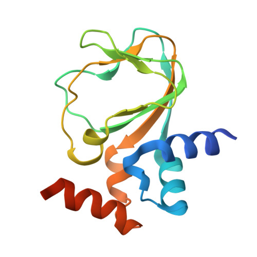
3CO2

Mlotik1 ion channel cyclic-nucleotide binding domain mutant


Experimental Data Snapshot

  • Method: X-RAY DIFFRACTION
  • Resolution: 2.90 Å
  • R-Value Free: 0.306 
  • R-Value Work: 0.261 
  • R-Value Observed: 0.263 

wwPDB Validation   3D Report Full Report


This is version 1.4 of the entry. See complete history


Literature

Structural and Energetic Analysis of Activation by a Cyclic Nucleotide Binding Domain.

Altieri, S.L.Clayton, G.M.Silverman, W.R.Olivares, A.O.De La Cruz, E.M.Thomas, L.R.Morais-Cabral, J.H.

(2008) J Mol Biol 381: 655-669

  • DOI: https://doi.org/10.1016/j.jmb.2008.06.011
  • Primary Citation of Related Structures:  
    3CL1, 3CLP, 3CO2

  • PubMed Abstract: 

    MlotiK1 is a prokaryotic homolog of cyclic-nucleotide-dependent ion channels that contains an intracellular C-terminal cyclic nucleotide binding (CNB) domain. X-ray structures of the CNB domain have been solved in the absence of ligand and bound to cAMP. Both the full-length channel and CNB domain fragment are easily expressed and purified, making MlotiK1 a useful model system for dissecting activation by ligand binding. We have used X-ray crystallography to determine three new MlotiK1 CNB domain structures: a second apo configuration, a cGMP-bound structure, and a second cAMP-bound structure. In combination, the five MlotiK1 CNB domain structures provide a unique opportunity for analyzing, within a single protein, the structural differences between the apo state and the bound state, and the structural variability within each state. With this analysis as a guide, we have probed the nucleotide selectivity and importance of specific residue side chains in ligand binding and channel activation. These data help to identify ligand-protein interactions that are important for ligand dependence in MlotiK1 and, more globally, in the class of nucleotide-dependent proteins.


  • Organizational Affiliation

    Department of Molecular Biophysics and Biochemistry, Yale University, New Haven, CT 06520-8114, USA.


Macromolecules
Find similar proteins by:  (by identity cutoff)  |  3D Structure
Entity ID: 1
MoleculeChains Sequence LengthOrganismDetailsImage
Mlotik1 ion channel protein
A, B, C, D
140Mesorhizobium lotiMutation(s): 1 
UniProt
Find proteins for Q98GN8 (Mesorhizobium japonicum (strain LMG 29417 / CECT 9101 / MAFF 303099))
Explore Q98GN8 
Go to UniProtKB:  Q98GN8
Entity Groups  
Sequence Clusters30% Identity50% Identity70% Identity90% Identity95% Identity100% Identity
UniProt GroupQ98GN8
Sequence Annotations
Expand
  • Reference Sequence
Experimental Data & Validation

Experimental Data

  • Method: X-RAY DIFFRACTION
  • Resolution: 2.90 Å
  • R-Value Free: 0.306 
  • R-Value Work: 0.261 
  • R-Value Observed: 0.263 
  • Space Group: P 21 21 2
Unit Cell:
Length ( Å )Angle ( ˚ )
a = 111.473α = 90
b = 132.349β = 90
c = 39.822γ = 90
Software Package:
Software NamePurpose
DENZOdata reduction
SCALEPACKdata scaling
REFMACrefinement
PDB_EXTRACTdata extraction

Structure Validation

View Full Validation Report


Entry History 

Deposition Data

Revision History  (Full details and data files)

  • Version 1.0: 2008-08-05
    Type: Initial release
  • Version 1.1: 2011-07-13
    Changes: Advisory, Version format compliance
  • Version 1.2: 2017-10-25
    Changes: Refinement description
  • Version 1.3: 2021-10-20
    Changes: Database references
  • Version 1.4: 2024-02-21
    Changes: Data collection