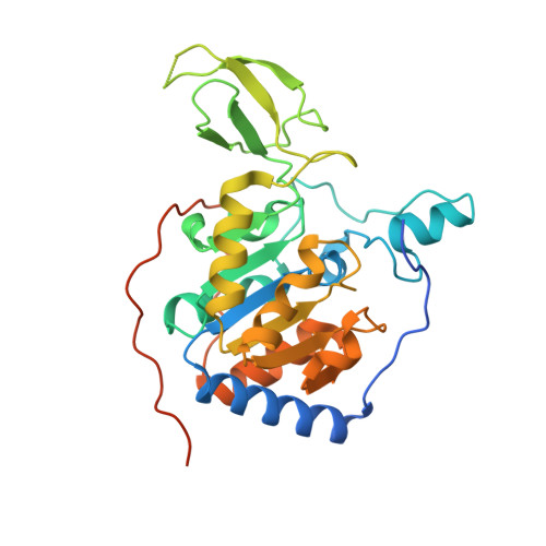
3K35

Crystal Structure of Human SIRT6


Experimental Data Snapshot

  • Method: X-RAY DIFFRACTION
  • Resolution: 2.00 Å
  • R-Value Free: 0.267 
  • R-Value Work: 0.202 
  • R-Value Observed: 0.202 

Starting Model: experimental
View more details

wwPDB Validation   3D Report Full Report


Ligand Structure Quality Assessment 


This is version 1.3 of the entry. See complete history


Literature

Structure and biochemical functions of SIRT6.

Pan, P.W.Feldman, J.L.Devries, M.K.Dong, A.Edwards, A.M.Denu, J.M.

(2011) J Biol Chem 286: 14575-14587

  • DOI: https://doi.org/10.1074/jbc.M111.218990
  • Primary Citation of Related Structures:  
    3K35, 3PKI, 3PKJ

  • PubMed Abstract: 

    SIRT6 is a member of the evolutionarily conserved sirtuin family of NAD(+)-dependent protein deacetylases and functions in genomic stability and transcriptional control of glucose metabolism. Early reports suggested that SIRT6 performs ADP-ribosylation, whereas more recent studies have suggested that SIRT6 functions mainly as a histone deacetylase. Thus, the molecular functions of SIRT6 remain uncertain. Here, we perform biochemical, kinetic, and structural studies to provide new mechanistic insight into the functions of SIRT6. Utilizing three different assays, we provide biochemical and kinetic evidence that SIRT6-dependent histone deacetylation produces O-acetyl-ADP-ribose but at a rate ∼1,000 times slower than other highly active sirtuins. To understand the molecular basis for such low deacetylase activity, we solved the first crystal structures of this class IV sirtuin in complex with ADP-ribose and the non-hydrolyzable analog of O-acetyl-ADP-ribose, 2'-N-acetyl-ADP-ribose. The structures revealed unique features of human SIRT6, including a splayed zinc-binding domain and the absence of a helix bundle that in other sirtuin structures connects the zinc-binding motif and Rossmann fold domain. SIRT6 also lacks the conserved, highly flexible, NAD(+)-binding loop and instead contains a stable single helix. These differences led us to hypothesize that SIRT6, unlike all other studied sirtuins, would be able to bind NAD(+) in the absence of an acetylated substrate. Indeed, we found that SIRT6 binds NAD(+) with relatively high affinity (K(d) = 27 ± 1 μM) in the absence of an acetylated substrate. Isothermal titration calorimetry and tryptophan fluorescence binding assays suggested that ADP-ribose and NAD(+) induce different structural perturbations and that NADH does not bind to SIRT6. Collectively, these new insights imply a unique activating mechanism and/or the possibility that SIRT6 could act as an NAD(+) metabolite sensor.


  • Organizational Affiliation

    Structural Genomics Consortium, University of Toronto, Toronto, Ontario M5G 1L7, Canada.


Macromolecules
Find similar proteins by:  (by identity cutoff)  |  3D Structure
Entity ID: 1
MoleculeChains Sequence LengthOrganismDetailsImage
NAD-dependent deacetylase sirtuin-6
A, B, C, D, E
A, B, C, D, E, F
318Homo sapiensMutation(s): 0 
Gene Names: SIR2L6SIRT6
EC: 3.5.1 (PDB Primary Data), 2.4.2 (UniProt), 2.3.1 (UniProt), 2.3.1.286 (UniProt)
UniProt & NIH Common Fund Data Resources
Find proteins for Q8N6T7 (Homo sapiens)
Explore Q8N6T7 
Go to UniProtKB:  Q8N6T7
PHAROS:  Q8N6T7
GTEx:  ENSG00000077463 
Entity Groups  
Sequence Clusters30% Identity50% Identity70% Identity90% Identity95% Identity100% Identity
UniProt GroupQ8N6T7
Sequence Annotations
Expand
  • Reference Sequence
Small Molecules
Ligands 4 Unique
IDChains Name / Formula / InChI Key2D Diagram3D Interactions
APR
Query on APR

Download Ideal Coordinates CCD File 
DA [auth D]
H [auth A]
KA [auth E]
Q [auth B]
RA [auth F]
DA [auth D],
H [auth A],
KA [auth E],
Q [auth B],
RA [auth F],
X [auth C]
ADENOSINE-5-DIPHOSPHORIBOSE
C15 H23 N5 O14 P2
SRNWOUGRCWSEMX-KEOHHSTQSA-N
SO4
Query on SO4

Download Ideal Coordinates CCD File 
EA [auth D]
FA [auth D]
I [auth A]
J [auth A]
K [auth A]
EA [auth D],
FA [auth D],
I [auth A],
J [auth A],
K [auth A],
L [auth A],
LA [auth E],
MA [auth E],
R [auth B],
S [auth B],
SA [auth F],
TA [auth F],
Y [auth C]
SULFATE ION
O4 S
QAOWNCQODCNURD-UHFFFAOYSA-L
ZN
Query on ZN

Download Ideal Coordinates CCD File 
CA [auth D]
G [auth A]
JA [auth E]
P [auth B]
QA [auth F]
CA [auth D],
G [auth A],
JA [auth E],
P [auth B],
QA [auth F],
W [auth C]
ZINC ION
Zn
PTFCDOFLOPIGGS-UHFFFAOYSA-N
UNX
Query on UNX

Download Ideal Coordinates CCD File 
AA [auth C]
BA [auth C]
GA [auth D]
HA [auth D]
IA [auth D]
AA [auth C],
BA [auth C],
GA [auth D],
HA [auth D],
IA [auth D],
M [auth A],
N [auth A],
NA [auth E],
O [auth A],
OA [auth E],
PA [auth E],
T [auth B],
U [auth B],
UA [auth F],
V [auth B],
VA [auth F],
WA [auth F],
Z [auth C]
UNKNOWN ATOM OR ION
X
Experimental Data & Validation

Experimental Data

  • Method: X-RAY DIFFRACTION
  • Resolution: 2.00 Å
  • R-Value Free: 0.267 
  • R-Value Work: 0.202 
  • R-Value Observed: 0.202 
  • Space Group: P 1 21 1
Unit Cell:
Length ( Å )Angle ( ˚ )
a = 89.388α = 90
b = 136.266β = 119.87
c = 89.26γ = 90
Software Package:
Software NamePurpose
HKL-2000data collection
PHASERphasing
REFMACrefinement
HKL-2000data reduction
HKL-2000data scaling

Structure Validation

View Full Validation Report


Ligand Structure Quality Assessment 


Entry History 

Revision History  (Full details and data files)

  • Version 1.0: 2009-12-08
    Type: Initial release
  • Version 1.1: 2011-07-13
    Changes: Version format compliance
  • Version 1.2: 2017-02-01
    Changes: Database references
  • Version 1.3: 2023-09-06
    Changes: Data collection, Database references, Derived calculations, Refinement description