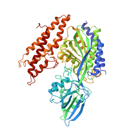
3KAS

Machupo virus GP1 bound to human transferrin receptor 1


Experimental Data Snapshot

  • Method: X-RAY DIFFRACTION
  • Resolution: 2.40 Å
  • R-Value Free: 0.238 
  • R-Value Work: 0.189 
  • R-Value Observed: 0.192 

Starting Model: experimental
View more details

wwPDB Validation   3D Report Full Report


Ligand Structure Quality Assessment 


This is version 2.2 of the entry. See complete history


Literature

Structural basis for receptor recognition by New World hemorrhagic fever arenaviruses.

Abraham, J.Corbett, K.D.Farzan, M.Choe, H.Harrison, S.C.

(2010) Nat Struct Mol Biol 17: 438-444

  • DOI: https://doi.org/10.1038/nsmb.1772
  • Primary Citation of Related Structures:  
    3KAS

  • PubMed Abstract: 

    New World hemorrhagic fever arenaviruses are rodent-borne agents that cause severe human disease. The GP1 subunit of the surface glycoprotein mediates cell attachment through transferrin receptor 1 (TfR1). We report the structure of Machupo virus (MACV) GP1 bound with human TfR1. Atomic details of the GP1-TfR1 interface clarify the importance of TfR1 residues implicated in New World arenavirus host specificity. Analysis of sequence variation among New World arenavirus GP1s and their host-species receptors, in light of the molecular structure, indicates determinants of viral zoonotic transmission. Infectivities of pseudoviruses in cells expressing mutated TfR1 confirm that contacts at the tip of the TfR1 apical domain determine the capacity of human TfR1 to mediate infection by particular New World arenaviruses. We propose that New World arenaviruses that are pathogenic to humans fortuitously acquired affinity for human TfR1 during adaptation to TfR1 of their natural hosts.


  • Organizational Affiliation

    Laboratory of Molecular Medicine, Harvard Medical School, Boston, Massachusetts, USA.


Macromolecules
Find similar proteins by:  (by identity cutoff)  |  3D Structure
Entity ID: 1
MoleculeChains Sequence LengthOrganismDetailsImage
Transferrin receptor protein 1640Homo sapiensMutation(s): 1 
UniProt & NIH Common Fund Data Resources
Find proteins for P02786 (Homo sapiens)
Explore P02786 
Go to UniProtKB:  P02786
PHAROS:  P02786
GTEx:  ENSG00000072274 
Entity Groups  
Sequence Clusters30% Identity50% Identity70% Identity90% Identity95% Identity100% Identity
UniProt GroupP02786
Glycosylation
Glycosylation Sites: 2Go to GlyGen: P02786-1
Sequence Annotations
Expand
  • Reference Sequence
Find similar proteins by:  (by identity cutoff)  |  3D Structure
Entity ID: 2
MoleculeChains Sequence LengthOrganismDetailsImage
Glycoprotein162Mammarenavirus machupoenseMutation(s): 0 
UniProt
Find proteins for Q8AZ57 (Machupo virus)
Explore Q8AZ57 
Go to UniProtKB:  Q8AZ57
Entity Groups  
Sequence Clusters30% Identity50% Identity70% Identity90% Identity95% Identity100% Identity
UniProt GroupQ8AZ57
Glycosylation
Glycosylation Sites: 4
Sequence Annotations
Expand
  • Reference Sequence
Oligosaccharides

Help

Entity ID: 3
MoleculeChains Length2D Diagram Glycosylation3D Interactions
2-acetamido-2-deoxy-beta-D-glucopyranose-(1-4)-[alpha-L-fucopyranose-(1-6)]2-acetamido-2-deoxy-beta-D-glucopyranose
C
3N-Glycosylation
Glycosylation Resources
GlyTouCan:  G21290RB
GlyCosmos:  G21290RB
GlyGen:  G21290RB
Entity ID: 4
MoleculeChains Length2D Diagram Glycosylation3D Interactions
alpha-D-mannopyranose-(1-3)-[alpha-D-mannopyranose-(1-6)]alpha-D-mannopyranose-(1-6)-[alpha-D-mannopyranose-(1-3)]beta-D-mannopyranose-(1-4)-2-acetamido-2-deoxy-beta-D-glucopyranose-(1-4)-2-acetamido-2-deoxy-beta-D-glucopyranose
D
7N-Glycosylation
Glycosylation Resources
GlyTouCan:  G55220VL
GlyCosmos:  G55220VL
GlyGen:  G55220VL
Small Molecules
Ligands 3 Unique
IDChains Name / Formula / InChI Key2D Diagram3D Interactions
NAG
Query on NAG

Download Ideal Coordinates CCD File 
E [auth A],
F [auth A],
L [auth B],
M [auth B]
2-acetamido-2-deoxy-beta-D-glucopyranose
C8 H15 N O6
OVRNDRQMDRJTHS-FMDGEEDCSA-N
PO4
Query on PO4

Download Ideal Coordinates CCD File 
G [auth A]
H [auth A]
I [auth A]
J [auth A]
N [auth B]
G [auth A],
H [auth A],
I [auth A],
J [auth A],
N [auth B],
O [auth B]
PHOSPHATE ION
O4 P
NBIIXXVUZAFLBC-UHFFFAOYSA-K
K
Query on K

Download Ideal Coordinates CCD File 
K [auth A]POTASSIUM ION
K
NPYPAHLBTDXSSS-UHFFFAOYSA-N
Experimental Data & Validation

Experimental Data

  • Method: X-RAY DIFFRACTION
  • Resolution: 2.40 Å
  • R-Value Free: 0.238 
  • R-Value Work: 0.189 
  • R-Value Observed: 0.192 
  • Space Group: C 2 2 21
Unit Cell:
Length ( Å )Angle ( ˚ )
a = 143.951α = 90
b = 170.228β = 90
c = 98.471γ = 90
Software Package:
Software NamePurpose
ADSCdata collection
PHASERphasing
PHENIXrefinement
HKL-2000data reduction
HKL-2000data scaling

Structure Validation

View Full Validation Report


Ligand Structure Quality Assessment 


Entry History 

Deposition Data

Revision History  (Full details and data files)

  • Version 1.0: 2010-03-09
    Type: Initial release
  • Version 1.1: 2011-07-13
    Changes: Version format compliance
  • Version 2.0: 2020-07-29
    Type: Remediation
    Reason: Carbohydrate remediation
    Changes: Advisory, Atomic model, Data collection, Database references, Derived calculations, Structure summary
  • Version 2.1: 2023-09-06
    Changes: Data collection, Database references, Refinement description, Structure summary
  • Version 2.2: 2024-10-09
    Changes: Structure summary