3KB6 | pdb_00003kb6

Crystal structure of D-Lactate dehydrogenase from aquifex aeolicus complexed with NAD and Lactic acid


Experimental Data Snapshot

  • Method: X-RAY DIFFRACTION
  • Resolution: 2.12 Å
  • R-Value Free: 
    0.212 (Depositor), 0.210 (DCC) 
  • R-Value Work: 
    0.164 (Depositor), 0.170 (DCC) 
  • R-Value Observed: 
    0.167 (Depositor) 

Starting Model: experimental
View more details

wwPDB Validation   3D Report Full Report


Ligand Structure Quality Assessment 

Created with Raphaël 2.3.0Worse 01 BetterLigand structure goodness of fit to experimental dataBest fitted NADClick on this verticalbar to view details

This is version 2.0 of the entry. See complete history


Literature

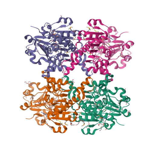
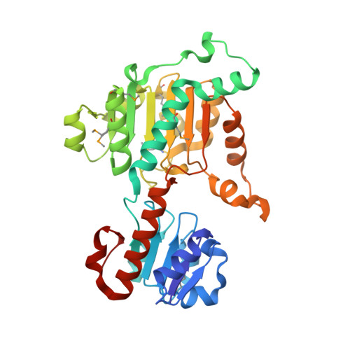
Structure of D-lactate dehydrogenase from Aquifex aeolicus complexed with NAD(+) and lactic acid (or pyruvate).

Antonyuk, S.V.Strange, R.W.Ellis, M.J.Bessho, Y.Kuramitsu, S.Inoue, Y.Yokoyama, S.Hasnain, S.S.

(2009) Acta Crystallogr Sect F Struct Biol Cryst Commun 65: 1209-1213

  • DOI: https://doi.org/10.1107/S1744309109044935
  • Primary Citation of Related Structures:  
    3KB6

  • PubMed Abstract: 

    The crystal structure of D-lactate dehydrogenase from Aquifex aeolicus (aq_727) was determined to 2.12 A resolution in space group P2(1)2(1)2(1), with unit-cell parameters a = 90.94, b = 94.43, c = 188.85 A. The structure was solved by molecular replacement using the coenzyme-binding domain of Lactobacillus helveticus D-lactate dehydrogenase and contained two homodimers in the asymmetric unit. Each subunit of the homodimer was found to be in a ;closed' conformation with the NADH cofactor bound to the coenzyme-binding domain and with a lactate (or pyruvate) molecule bound at the interdomain active-site cleft.


  • Organizational Affiliation

    Molecular Biophysics Group, School of Biological Sciences, University of Liverpool, Crown Street, Liverpool L69 7ZB, England.


Macromolecules
Find similar proteins by:  (by identity cutoff)  |  3D Structure
Entity ID: 1
MoleculeChains Sequence LengthOrganismDetailsImage
D-lactate dehydrogenase
A, B, C, D
334Aquifex aeolicusMutation(s): 0 
Gene Names: aq_727ldhA
EC: 1.1.1.28
UniProt
Find proteins for O66939 (Aquifex aeolicus (strain VF5))
Explore O66939 
Go to UniProtKB:  O66939
Entity Groups  
Sequence Clusters30% Identity50% Identity70% Identity90% Identity95% Identity100% Identity
UniProt GroupO66939
Sequence Annotations
Expand
  • Reference Sequence
Small Molecules
Ligands 6 Unique
IDChains Name / Formula / InChI Key2D Diagram3D Interactions
NAD
Query on NAD

Download Ideal Coordinates CCD File 
G [auth A],
JA [auth C],
QA [auth D],
Y [auth B]
NICOTINAMIDE-ADENINE-DINUCLEOTIDE
C21 H27 N7 O14 P2
BAWFJGJZGIEFAR-NNYOXOHSSA-N
1PE
Query on 1PE

Download Ideal Coordinates CCD File 
AA [auth B]
BA [auth B]
FA [auth B]
I [auth A]
L [auth A]
PENTAETHYLENE GLYCOL
C10 H22 O6
JLFNLZLINWHATN-UHFFFAOYSA-N
PEG
Query on PEG

Download Ideal Coordinates CCD File 
GA [auth C]
H [auth A]
K [auth A]
M [auth A]
NA [auth C]
DI(HYDROXYETHYL)ETHER
C4 H10 O3
MTHSVFCYNBDYFN-UHFFFAOYSA-N
GOL
Query on GOL

Download Ideal Coordinates CCD File 
CA [auth B]
DA [auth B]
EA [auth B]
KA [auth C]
MA [auth C]
GLYCEROL
C3 H8 O3
PEDCQBHIVMGVHV-UHFFFAOYSA-N
LAC
Query on LAC

Download Ideal Coordinates CCD File 
F [auth A],
IA [auth C],
PA [auth D],
X [auth B]
LACTIC ACID
C3 H6 O3
JVTAAEKCZFNVCJ-UWTATZPHSA-N
PPI
Query on PPI

Download Ideal Coordinates CCD File 
E [auth A],
HA [auth C],
J [auth A],
OA [auth D],
W [auth B]
PROPANOIC ACID
C3 H6 O2
XBDQKXXYIPTUBI-UHFFFAOYSA-N
Modified Residues  1 Unique
IDChains TypeFormula2D DiagramParent
MSE
Query on MSE
A, B, C, D
L-PEPTIDE LINKINGC5 H11 N O2 SeMET
Experimental Data & Validation

Experimental Data

  • Method: X-RAY DIFFRACTION
  • Resolution: 2.12 Å
  • R-Value Free:  0.212 (Depositor), 0.210 (DCC) 
  • R-Value Work:  0.164 (Depositor), 0.170 (DCC) 
  • R-Value Observed: 0.167 (Depositor) 
Space Group: P 21 21 21
Unit Cell:
Length ( Å )Angle ( ˚ )
a = 90.938α = 90
b = 94.432β = 90
c = 188.849γ = 90
Software Package:
Software NamePurpose
DENZOdata reduction
SCALEPACKdata scaling
MOLREPphasing
REFMACrefinement
PDB_EXTRACTdata extraction
HKL-2000data collection

Structure Validation

View Full Validation Report


Ligand Structure Quality Assessment 

Created with Raphaël 2.3.0Worse 01 BetterLigand structure goodness of fit to experimental dataBest fitted NADClick on this verticalbar to view details

Entry History 

Deposition Data

Revision History  (Full details and data files)

  • Version 1.0: 2009-11-10
    Type: Initial release
  • Version 1.1: 2011-07-13
    Changes: Non-polymer description, Version format compliance
  • Version 1.2: 2017-11-01
    Changes: Advisory, Refinement description
  • Version 1.3: 2023-11-01
    Changes: Advisory, Data collection, Database references, Derived calculations, Refinement description
  • Version 2.0: 2023-11-15
    Changes: Atomic model, Data collection