3QEP | pdb_00003qep

RB69 DNA Polymerase (L561A/S565G/Y567A) Ternary Complex with dTTP Opposite Difluorotoluene Nucleoside


Experimental Data Snapshot

  • Method: X-RAY DIFFRACTION
  • Resolution: 1.80 Å
  • R-Value Free: 
    0.204 (Depositor), 0.210 (DCC) 
  • R-Value Work: 
    0.167 (Depositor), 0.190 (DCC) 
  • R-Value Observed: 
    0.169 (Depositor) 

wwPDB Validation   3D Report Full Report


Ligand Structure Quality Assessment 

Created with Raphaël 2.3.0Worse 01 BetterLigand structure goodness of fit to experimental dataBest fitted TTPClick on this verticalbar to view details

This is version 1.1 of the entry. See complete history


Literature

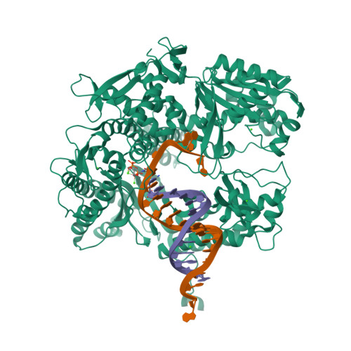
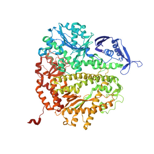
Hydrogen-bonding capability of a templating difluorotoluene nucleotide residue in an RB69 DNA polymerase ternary complex.

Xia, S.Konigsberg, W.H.Wang, J.

(2011) J Am Chem Soc 133: 10003-10005

  • DOI: https://doi.org/10.1021/ja2021735
  • Primary Citation of Related Structures:  
    3QEP, 3RWU

  • PubMed Abstract: 

    Results obtained using 2,4-difluorotoluene nucleobase (dF) as a nonpolar thymine isostere by Kool and colleagues challenged the Watson-Crick dogma that hydrogen bonds between complementary bases are an absolute requirement for accurate DNA replication. Here, we report crystal structure of an RB69 DNA polymerase L561A/S565G/Y567A triple mutant ternary complex with a templating dF opposite dTTP at 1.8 Å-resolution. In this structure, direct hydrogen bonds were observed between: (i) dF and the incoming dTTP, (ii) dF and residue G568 of the polymerase, and (iii) dF and ordered water molecules surrounding the nascent base pair. Therefore, this structure provides evidence that a templating dF can form novel hydrogen bonds with the incoming dTTP and with the enzyme that differ from those formed with a templating dT.


  • Organizational Affiliation

    Department of Molecular Biophysics and Biochemistry, Yale University, New Haven, Connecticut 06520, USA.


Macromolecules

Find similar proteins by:  (by identity cutoff)  |  3D Structure
Entity ID: 1
MoleculeChains Sequence LengthOrganismDetailsImage
DNA polymerase903Escherichia phage RB69Mutation(s): 3 
Gene Names: 43
EC: 2.7.7.7 (PDB Primary Data), 3.1.11 (UniProt)
UniProt
Find proteins for Q38087 (Escherichia phage RB69)
Explore Q38087 
Go to UniProtKB:  Q38087
Entity Groups  
Sequence Clusters30% Identity50% Identity70% Identity90% Identity95% Identity100% Identity
UniProt GroupQ38087
Sequence Annotations
Expand
  • Reference Sequence
Small Molecules
Ligands 2 Unique
IDChains Name / Formula / InChI Key2D Diagram3D Interactions
TTP
Query on TTP

Download Ideal Coordinates CCD File 
D [auth A]THYMIDINE-5'-TRIPHOSPHATE
C10 H17 N2 O14 P3
NHVNXKFIZYSCEB-XLPZGREQSA-N
CA
Query on CA

Download Ideal Coordinates CCD File 
E [auth A]
F [auth A]
G [auth A]
H [auth A]
I [auth A]
CALCIUM ION
Ca
BHPQYMZQTOCNFJ-UHFFFAOYSA-N
Experimental Data & Validation

Experimental Data

  • Method: X-RAY DIFFRACTION
  • Resolution: 1.80 Å
  • R-Value Free:  0.204 (Depositor), 0.210 (DCC) 
  • R-Value Work:  0.167 (Depositor), 0.190 (DCC) 
  • R-Value Observed: 0.169 (Depositor) 
Space Group: P 21 21 21
Unit Cell:
Length ( Å )Angle ( ˚ )
a = 75.133α = 90
b = 119.774β = 90
c = 130.481γ = 90
Software Package:
Software NamePurpose
HKL-2000data collection
AMoREphasing
REFMACrefinement
HKL-2000data reduction
HKL-2000data scaling

Structure Validation

View Full Validation Report


Ligand Structure Quality Assessment 

Created with Raphaël 2.3.0Worse 01 BetterLigand structure goodness of fit to experimental dataBest fitted TTPClick on this verticalbar to view details

Entry History 

Deposition Data

Revision History  (Full details and data files)

  • Version 1.0: 2011-08-31
    Type: Initial release
  • Version 1.1: 2024-02-21
    Changes: Data collection, Database references, Derived calculations