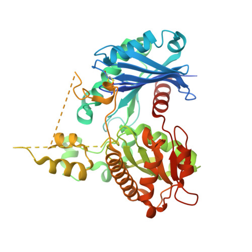
Structural and mechanistic investigations on Salmonella typhimurium acetate kinase (AckA): identification of a putative ligand binding pocket at the dimeric interface
Chittori, S., Savithri, H.S., Murthy, M.R.N.(2012) BMC Struct Biol 12: 24-24
- PubMed: 23031654
- DOI: https://doi.org/10.1186/1472-6807-12-24
- Primary Citation of Related Structures:
3SK3, 3SLC - PubMed Abstract:
Bacteria such as Escherichia coli and Salmonella typhimurium can utilize acetate as the sole source of carbon and energy. Acetate kinase (AckA) and phosphotransacetylase (Pta), key enzymes of acetate utilization pathway, regulate flux of metabolites in glycolysis, gluconeogenesis, TCA cycle, glyoxylate bypass and fatty acid metabolism. Here we report kinetic characterization of S. typhimurium AckA (StAckA) and structures of its unliganded (Form-I, 2.70 Å resolution) and citrate-bound (Form-II, 1.90 Å resolution) forms. The enzyme showed broad substrate specificity with k(cat)/K(m) in the order of acetate > propionate > formate. Further, the Km for acetyl-phosphate was significantly lower than for acetate and the enzyme could catalyze the reverse reaction (i.e. ATP synthesis) more efficiently. ATP and Mg(2+) could be substituted by other nucleoside 5'-triphosphates (GTP, UTP and CTP) and divalent cations (Mn(2+) and Co(2+)), respectively. Form-I StAckA represents the first structural report of an unliganded AckA. StAckA protomer consists of two domains with characteristic βββαβαβα topology of ASKHA superfamily of proteins. These domains adopt an intermediate conformation compared to that of open and closed forms of ligand-bound Methanosarcina thermophila AckA (MtAckA). Spectroscopic and structural analyses of StAckA further suggested occurrence of inter-domain motion upon ligand-binding. Unexpectedly, Form-II StAckA structure showed a drastic change in the conformation of residues 230-300 compared to that of Form-I. Further investigation revealed electron density corresponding to a citrate molecule in a pocket located at the dimeric interface of Form-II StAckA. Interestingly, a similar dimeric interface pocket lined with largely conserved residues could be identified in Form-I StAckA as well as in other enzymes homologous to AckA suggesting that ligand binding at this pocket may influence the function of these enzymes. The biochemical and structural characterization of StAckA reported here provides insights into the biochemical specificity, overall fold, thermal stability, molecular basis of ligand binding and inter-domain motion in AckA family of enzymes. Dramatic conformational differences observed between unliganded and citrate-bound forms of StAckA led to identification of a putative ligand-binding pocket at the dimeric interface of StAckA with implications for enzymatic function.
Organizational Affiliation:
Molecular Biophysics Unit, Indian Institute of Science, Bangalore, Karnataka 560012, India.


