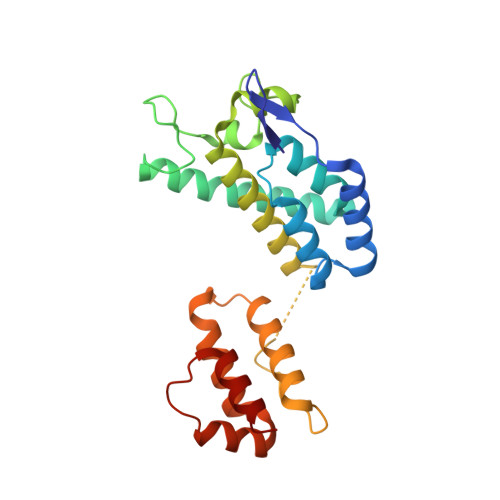
A structural model for the generation of continuous curvature on the surface of a retroviral capsid.
Bailey, G.D., Hyun, J.K., Mitra, A.K., Kingston, R.L.(2012) J Mol Biol 417: 212-223
- PubMed: 22306463
- DOI: https://doi.org/10.1016/j.jmb.2012.01.014
- Primary Citation of Related Structures:
3TIR - PubMed Abstract:
The genome of a retrovirus is surrounded by a convex protein shell, or capsid, that helps facilitate infection. The major part of the capsid surface is formed by interlocking capsid protein (CA) hexamers. We report electron and X-ray crystallographic analysis of a variety of specimens assembled in vitro from Rous sarcoma virus (RSV) CA. These specimens all contain CA hexamers arranged in planar layers, modeling the authentic capsid surface. The specimens differ only in the number of layers incorporated and in the disposition of each layer with respect to its neighbor. The body of each hexamer, formed by the N-terminal domain of CA, is connected to neighboring hexamers through C-terminal domain dimerization. The resulting layer structure is very malleable due to inter-domain flexibility. A helix-capping hydrogen bond between the two domains of RSV CA creates a pivot point, which is central to controlling their relative movement. A similar mechanism for the governance of inter-domain motion was recently described for the human immunodeficiency virus type 1 (HIV-1) capsid, although there is negligible sequence identity between RSV and HIV-1 CA in the region of contact, and the amino acids involved in creating the pivot are not conserved. Our observations allow development of a physically realistic model for the way neighboring hexamers can tilt out of plane, deforming the hexamer layer and generating the continuously curved surfaces that are a feature of all retroviral capsids.
Organizational Affiliation:
OncImmune Ltd, City Hospital, Nottingham NG5 1PB, UK.


