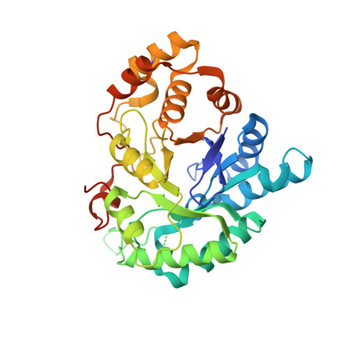
3UWE

AKR1C3 complexed with 3-phenoxybenzoic acid


Experimental Data Snapshot

  • Method: X-RAY DIFFRACTION
  • Resolution: 1.68 Å
  • R-Value Free: 0.214 
  • R-Value Work: 0.184 
  • R-Value Observed: 0.185 

Starting Model: experimental
View more details

wwPDB Validation   3D Report Full Report


Ligand Structure Quality Assessment 


This is version 1.2 of the entry. See complete history


Literature

Structure of AKR1C3 with 3-phenoxybenzoic acid bound

Jackson, V.J.Yosaatmadja, Y.Flanagan, J.U.Squire, C.J.

(2012) Acta Crystallogr Sect F Struct Biol Cryst Commun 68: 409-413

  • DOI: https://doi.org/10.1107/S1744309112009049
  • Primary Citation of Related Structures:  
    3UWE

  • PubMed Abstract: 

    Aldo-keto reductase 1C3 (AKR1C3) is a human enzyme that catalyzes the NADPH-dependent reduction of steroids and prostaglandins. AKR1C3 overexpression is associated with the proliferation of hormone-dependent cancers, most notably breast and prostate cancers. Nonsteroidal anti-inflammatory drugs (NSAIDs) and their analogues are well characterized inhibitors of AKR1C3. Here, the X-ray crystal structure of 3-phenoxybenzoic acid in complex with AKR1C3 is presented. This structure provides useful information for the future development of new anticancer agents by structure-guided drug design.


  • Organizational Affiliation

    School of Biological Sciences, University of Auckland, Private Bag 92019, Victoria St. West, Auckland, New Zealand.


Macromolecules
Find similar proteins by:  (by identity cutoff)  |  3D Structure
Entity ID: 1
MoleculeChains Sequence LengthOrganismDetailsImage
Aldo-keto reductase family 1 member C3331Homo sapiensMutation(s): 0 
Gene Names: AKR1C3DDH1HSD17B5KIAA0119PGFS
EC: 1.1.1.213 (PDB Primary Data), 1.1.1.112 (PDB Primary Data), 1.1.1.188 (PDB Primary Data), 1.1.1.63 (PDB Primary Data), 1.1.1.64 (PDB Primary Data), 1.3.1.20 (PDB Primary Data), 1.1.1.53 (UniProt), 1.1.1.62 (UniProt), 1.1.1.357 (UniProt), 1.1.1.239 (UniProt), 1.1.1 (UniProt), 1.1.1.210 (UniProt)
UniProt & NIH Common Fund Data Resources
Find proteins for P42330 (Homo sapiens)
Explore P42330 
Go to UniProtKB:  P42330
PHAROS:  P42330
GTEx:  ENSG00000196139 
Entity Groups  
Sequence Clusters30% Identity50% Identity70% Identity90% Identity95% Identity100% Identity
UniProt GroupP42330
Sequence Annotations
Expand
  • Reference Sequence
Small Molecules
Ligands 3 Unique
IDChains Name / Formula / InChI Key2D Diagram3D Interactions
NAP
Query on NAP

Download Ideal Coordinates CCD File 
B [auth A]NADP NICOTINAMIDE-ADENINE-DINUCLEOTIDE PHOSPHATE
C21 H28 N7 O17 P3
XJLXINKUBYWONI-NNYOXOHSSA-N
VJJ
Query on VJJ

Download Ideal Coordinates CCD File 
C [auth A]3-phenoxybenzoic acid
C13 H10 O3
NXTDJHZGHOFSQG-UHFFFAOYSA-N
EDO
Query on EDO

Download Ideal Coordinates CCD File 
D [auth A],
E [auth A],
F [auth A],
G [auth A]
1,2-ETHANEDIOL
C2 H6 O2
LYCAIKOWRPUZTN-UHFFFAOYSA-N
Modified Residues  1 Unique
IDChains TypeFormula2D DiagramParent
CSX
Query on CSX
A
L-PEPTIDE LINKINGC3 H7 N O3 SCYS
Binding Affinity Annotations 
IDSourceBinding Affinity
VJJ BindingDB:  3UWE IC50: 680 (nM) from 1 assay(s)
Experimental Data & Validation

Experimental Data

  • Method: X-RAY DIFFRACTION
  • Resolution: 1.68 Å
  • R-Value Free: 0.214 
  • R-Value Work: 0.184 
  • R-Value Observed: 0.185 
  • Space Group: P 21 21 21
Unit Cell:
Length ( Å )Angle ( ˚ )
a = 58.473α = 90
b = 64.699β = 90
c = 96.959γ = 90
Software Package:
Software NamePurpose
MAR345dtbdata collection
PHASERphasing
REFMACrefinement
XDSdata reduction
SCALAdata scaling

Structure Validation

View Full Validation Report


Ligand Structure Quality Assessment 


Entry History 

Deposition Data

Revision History  (Full details and data files)

  • Version 1.0: 2012-04-04
    Type: Initial release
  • Version 1.1: 2014-03-05
    Changes: Database references
  • Version 1.2: 2023-11-08
    Changes: Data collection, Database references, Derived calculations, Refinement description