3W5N

Crystal Structure of Streptomyces avermitilis alpha-L-rhamnosidase complexed with L-rhamnose


Experimental Data Snapshot

  • Method: X-RAY DIFFRACTION
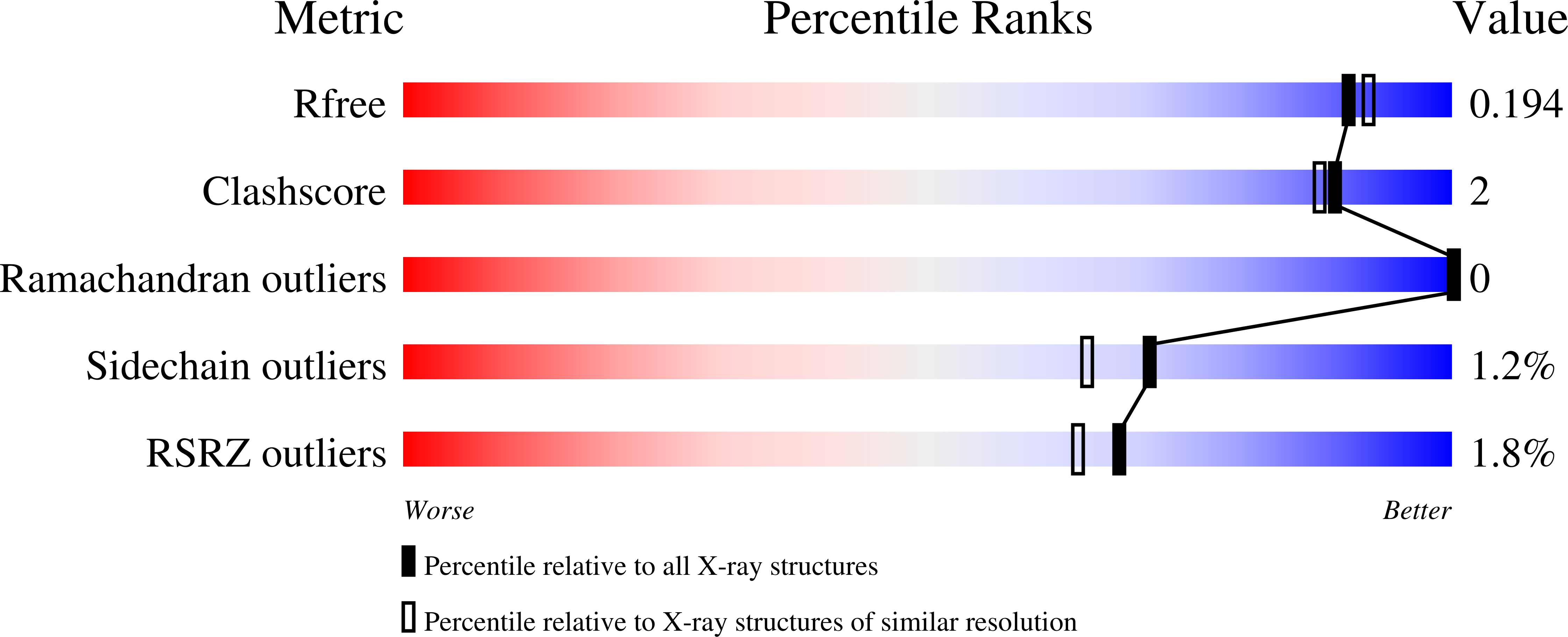
  • Resolution: 1.80 Å
  • R-Value Free: 0.194 
  • R-Value Work: 0.163 
  • R-Value Observed: 0.164 

Starting Model: experimental
View more details

wwPDB Validation   3D Report Full Report


Ligand Structure Quality Assessment 


This is version 1.4 of the entry. See complete history


Literature

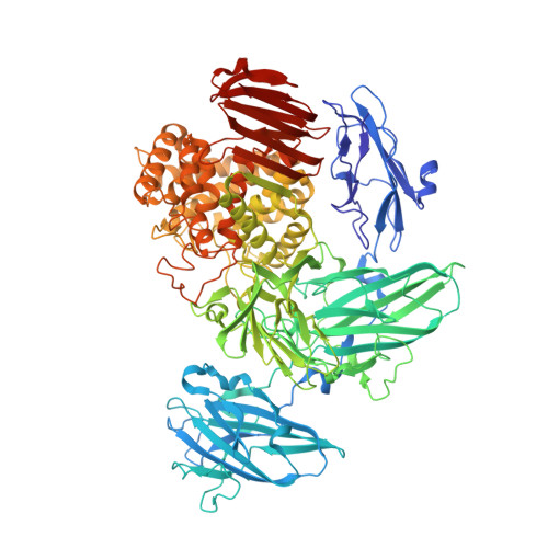
The structure of a Streptomyces avermitilis alpha-L-rhamnosidase reveals a novel carbohydrate-binding module CBM67 within the six-domain arrangement.

Fujimoto, Z.Jackson, A.Michikawa, M.Maehara, T.Momma, M.Henrissat, B.F.Gilbert, H.J.Kaneko, S.

(2013) J Biol Chem 288: 12376-12385

  • DOI: https://doi.org/10.1074/jbc.M113.460097
  • Primary Citation of Related Structures:  
    3W5M, 3W5N

  • PubMed Abstract: 

    α-L-rhamnosidases hydrolyze α-linked L-rhamnosides from oligosaccharides or polysaccharides. We determined the crystal structure of the glycoside hydrolase family 78 Streptomyces avermitilis α-L-rhamnosidase (SaRha78A) in its free and L-rhamnose complexed forms, which revealed the presence of six domains N, D, E, F, A, and C. In the ligand complex, L-rhamnose was bound in the proposed active site of the catalytic module, revealing the likely catalytic mechanism of SaRha78A. Glu(636) is predicted to donate protons to the glycosidic oxygen, and Glu(895) is the likely catalytic general base, activating the nucleophilic water, indicating that the enzyme operates through an inverting mechanism. Replacement of Glu(636) and Glu(895) resulted in significant loss of α-rhamnosidase activity. Domain D also bound L-rhamnose in a calcium-dependent manner, with a KD of 135 μm. Domain D is thus a non-catalytic carbohydrate binding module (designated SaCBM67). Mutagenesis and structural data identified the amino acids in SaCBM67 that target the features of L-rhamnose that distinguishes it from the other major sugars present in plant cell walls. Inactivation of SaCBM67 caused a substantial reduction in the activity of SaRha78A against the polysaccharide composite gum arabic, but not against aryl rhamnosides, indicating that SaCBM67 contributes to enzyme function against insoluble substrates.


  • Organizational Affiliation

    Biomolecular Research Unit, National Institute of Agrobiological Sciences, 2-1-2 Kannondai, Tsukuba 305-8602, Japan. zui@affrc.go.jp


Macromolecules
Find similar proteins by:  (by identity cutoff)  |  3D Structure
Entity ID: 1
MoleculeChains Sequence LengthOrganismDetailsImage
Putative rhamnosidase1,043Streptomyces avermitilis MA-4680 = NBRC 14893Mutation(s): 0 
Gene Names: SAV_828
EC: 3.2.1.40
UniProt
Find proteins for Q82PP4 (Streptomyces avermitilis (strain ATCC 31267 / DSM 46492 / JCM 5070 / NBRC 14893 / NCIMB 12804 / NRRL 8165 / MA-4680))
Explore Q82PP4 
Go to UniProtKB:  Q82PP4
Entity Groups  
Sequence Clusters30% Identity50% Identity70% Identity90% Identity95% Identity100% Identity
UniProt GroupQ82PP4
Sequence Annotations
Expand
  • Reference Sequence
Small Molecules
Ligands 4 Unique
IDChains Name / Formula / InChI Key2D Diagram3D Interactions
RAM
Query on RAM

Download Ideal Coordinates CCD File 
C [auth A],
D [auth A],
E [auth A],
F [auth A],
G [auth A]
alpha-L-rhamnopyranose
C6 H12 O5
SHZGCJCMOBCMKK-HGVZOGFYSA-N
MPD
Query on MPD

Download Ideal Coordinates CCD File 
H [auth A],
I [auth A],
J [auth A]
(4S)-2-METHYL-2,4-PENTANEDIOL
C6 H14 O2
SVTBMSDMJJWYQN-YFKPBYRVSA-N
CA
Query on CA

Download Ideal Coordinates CCD File 
B [auth A]CALCIUM ION
Ca
BHPQYMZQTOCNFJ-UHFFFAOYSA-N
NA
Query on NA

Download Ideal Coordinates CCD File 
K [auth A],
L [auth A],
M [auth A]
SODIUM ION
Na
FKNQFGJONOIPTF-UHFFFAOYSA-N
Binding Affinity Annotations 
IDSourceBinding Affinity
RAM PDBBind:  3W5N Kd: 1.35e+5 (nM) from 1 assay(s)
Experimental Data & Validation

Experimental Data

  • Method: X-RAY DIFFRACTION
  • Resolution: 1.80 Å
  • R-Value Free: 0.194 
  • R-Value Work: 0.163 
  • R-Value Observed: 0.164 
  • Space Group: P 1 21 1
Unit Cell:
Length ( Å )Angle ( ˚ )
a = 53.063α = 90
b = 128.559β = 99.85
c = 75.26γ = 90
Software Package:
Software NamePurpose
SERGUIdata collection
REFMACrefinement
HKL-2000data reduction
SCALEPACKdata scaling
REFMACphasing

Structure Validation

View Full Validation Report


Ligand Structure Quality Assessment 


Entry History 

Deposition Data

Revision History  (Full details and data files)

  • Version 1.0: 2013-03-20
    Type: Initial release
  • Version 1.1: 2013-10-16
    Changes: Database references
  • Version 1.2: 2017-11-22
    Changes: Refinement description
  • Version 1.3: 2020-07-29
    Type: Remediation
    Reason: Carbohydrate remediation
    Changes: Data collection, Database references, Derived calculations, Structure summary
  • Version 1.4: 2023-11-08
    Changes: Data collection, Database references, Refinement description, Structure summary