3ZX2

NTPDase1 in complex with Decavanadate


Experimental Data Snapshot

  • Method: X-RAY DIFFRACTION
  • Resolution: 1.81 Å
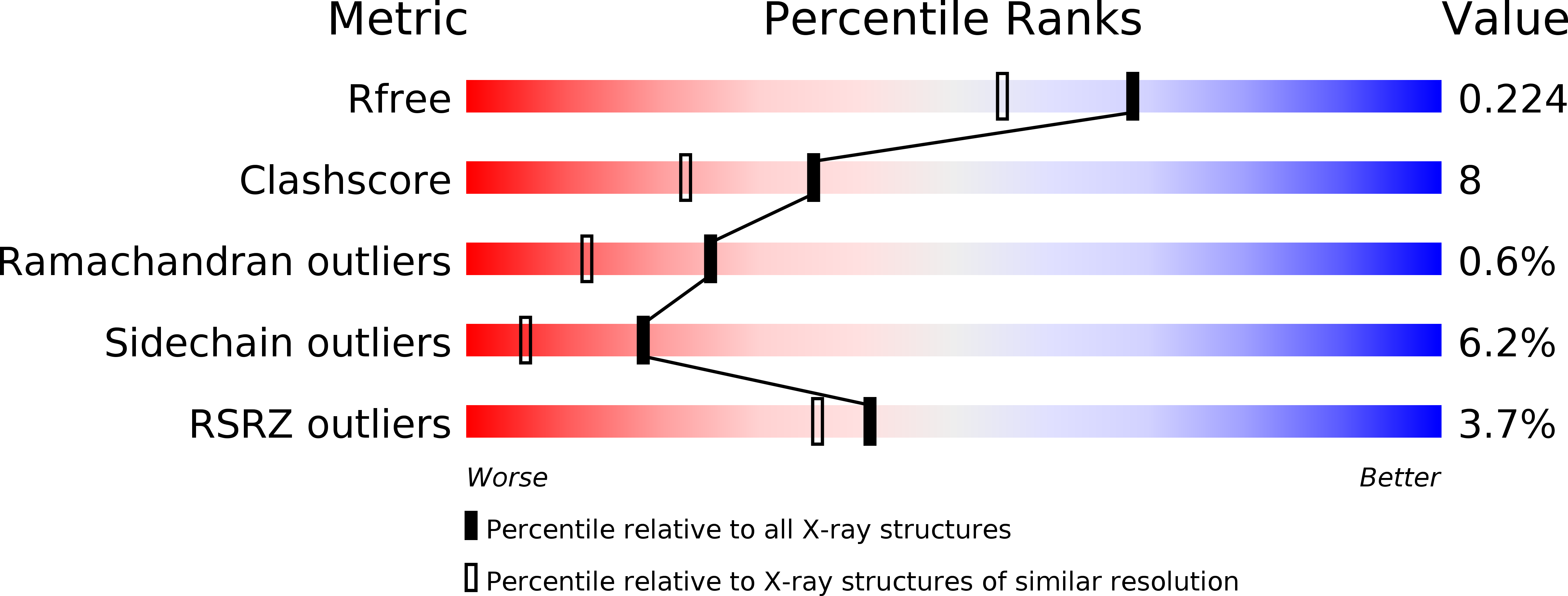
  • R-Value Free: 0.224 
  • R-Value Work: 0.175 
  • R-Value Observed: 0.176 

wwPDB Validation   3D Report Full Report


This is version 2.0 of the entry. See complete history


Literature

Crystallographic Evidence for a Domain Motion in Rat Nucleoside Triphosphate Diphosphohydrolase (Ntpdase) 1.

Zebisch, M.Krauss, M.Schafer, P.Strater, N.

(2012) J Mol Biol 415: 288

  • DOI: https://doi.org/10.1016/j.jmb.2011.10.050
  • Primary Citation of Related Structures:  
    3ZX0, 3ZX2, 3ZX3

  • PubMed Abstract: 

    Nucleoside triphosphate diphosphohydrolases (NTPDases) are a physiologically important class of membrane-bound ectonucleotidases responsible for the regulation of extracellular levels of nucleotides. CD39 or NTPDase1 is the dominant NTPDase of the vasculature. By hydrolyzing proinflammatory ATP and platelet-activating ADP to AMP, it blocks platelet aggregation and supports blood flow. Thus, great interest exists in understanding the structure and dynamics of this prototype member of the eukaryotic NTPDase family. Here, we report the crystal structure of a variant of soluble NTPDase1 lacking a putative membrane interaction loop identified between the two lobes of the catalytic domain. ATPase and ADPase activities of this variant are determined via a newly established kinetic isothermal titration calorimetry assay and compared to that of the soluble NTPDase1 variant characterized previously. Complex structures with decavanadate and heptamolybdate show that both polyoxometallates bind electrostatically to a loop that is involved in binding of the nucleobase. In addition, a comparison of the domain orientations of the four independent proteins in the crystal asymmetric unit provides the first direct experimental evidence for a domain motion of NTPDases. An interdomain rotation angle of up to 7.4° affects the active site cleft between the two lobes of the protein. Comparison with a previously solved bacterial NTPDase structure indicates that the domains may undergo relative rotational movements of more than 20°. Our data support the idea that the influence of transmembrane helix dynamics on activity is achieved by coupling to a domain motion.


  • Organizational Affiliation

    Institute of Bioanalytical Chemistry, Center for Biotechnology and Biomedicine, Faculty of Chemistry and Mineralogy, University of Leipzig, Deutscher Platz 5, 04103 Leipzig, Germany.


Macromolecules
Find similar proteins by:  (by identity cutoff)  |  3D Structure
Entity ID: 1
MoleculeChains Sequence LengthOrganismDetailsImage
ECTONUCLEOSIDE TRIPHOSPHATE DIPHOSPHOHYDROLASE 1
A, B, C, D
452Rattus norvegicusMutation(s): 0 
EC: 3.6.1.5
UniProt
Find proteins for P97687 (Rattus norvegicus)
Explore P97687 
Go to UniProtKB:  P97687
Entity Groups  
Sequence Clusters30% Identity50% Identity70% Identity90% Identity95% Identity100% Identity
UniProt GroupP97687
Sequence Annotations
Expand
  • Reference Sequence
Small Molecules
Ligands 4 Unique
IDChains Name / Formula / InChI Key2D Diagram3D Interactions
DVT
Query on DVT

Download Ideal Coordinates CCD File 
K [auth A],
RA [auth D],
T [auth B],
U [auth C]
DECAVANADATE
O28 V10
WOYKCMUEHQAMTC-UHFFFAOYSA-N
ACY
Query on ACY

Download Ideal Coordinates CCD File 
AA [auth C]
BA [auth C]
J [auth A]
MA [auth D]
NA [auth D]
AA [auth C],
BA [auth C],
J [auth A],
MA [auth D],
NA [auth D],
Q [auth B],
R [auth B]
ACETIC ACID
C2 H4 O2
QTBSBXVTEAMEQO-UHFFFAOYSA-N
CL
Query on CL

Download Ideal Coordinates CCD File 
E [auth A]
EA [auth D]
F [auth A]
FA [auth D]
G [auth A]
E [auth A],
EA [auth D],
F [auth A],
FA [auth D],
G [auth A],
GA [auth D],
H [auth A],
HA [auth D],
I [auth A],
IA [auth D],
JA [auth D],
KA [auth D],
L [auth B],
LA [auth D],
M [auth B],
N [auth B],
O [auth B],
P [auth B],
V [auth C],
W [auth C],
X [auth C],
Y [auth C],
Z [auth C]
CHLORIDE ION
Cl
VEXZGXHMUGYJMC-UHFFFAOYSA-M
NA
Query on NA

Download Ideal Coordinates CCD File 
CA [auth C]
DA [auth C]
OA [auth D]
PA [auth D]
QA [auth D]
CA [auth C],
DA [auth C],
OA [auth D],
PA [auth D],
QA [auth D],
S [auth B]
SODIUM ION
Na
FKNQFGJONOIPTF-UHFFFAOYSA-N
Experimental Data & Validation

Experimental Data

  • Method: X-RAY DIFFRACTION
  • Resolution: 1.81 Å
  • R-Value Free: 0.224 
  • R-Value Work: 0.175 
  • R-Value Observed: 0.176 
  • Space Group: C 1 2 1
Unit Cell:
Length ( Å )Angle ( ˚ )
a = 162.92α = 90
b = 80.985β = 117.49
c = 165.517γ = 90
Software Package:
Software NamePurpose
REFMACrefinement
XDSdata reduction
SCALAdata scaling

Structure Validation

View Full Validation Report


Entry History 

Deposition Data

Revision History  (Full details and data files)

  • Version 1.0: 2011-11-30
    Type: Initial release
  • Version 1.1: 2012-01-25
    Changes: Other
  • Version 1.2: 2018-01-31
    Changes: Source and taxonomy
  • Version 2.0: 2024-10-09
    Changes: Atomic model, Data collection, Database references, Derived calculations, Other, Structure summary