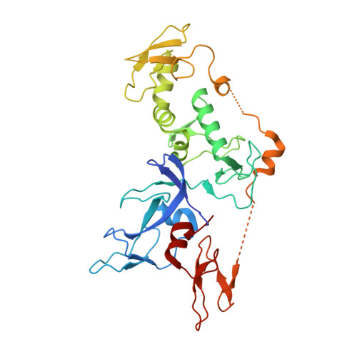
4BM9

Structure of the autoinhibited Parkin catalytic domain


Experimental Data Snapshot

  • Method: X-RAY DIFFRACTION
  • Resolution: 2.25 Å
  • R-Value Free: 0.218 
  • R-Value Work: 0.190 
  • R-Value Observed: 0.192 

wwPDB Validation   3D Report Full Report


This is version 1.2 of the entry. See complete history


Literature

Structure of the Human Parkin Ligase Domain in an Autoinhibited State.

Wauer, T.Komander, D.

(2013) EMBO J 32: 2099

  • DOI: https://doi.org/10.1038/emboj.2013.125
  • Primary Citation of Related Structures:  
    4BM9

  • PubMed Abstract: 

    Mutations in the protein Parkin are associated with Parkinson's disease (PD), the second most common neurodegenerative disease in men. Parkin is an E3 ubiquitin (Ub) ligase of the structurally uncharacterized RING-in-between-RING(IBR)-RING (RBR) family, which, in an HECT-like fashion, forms a catalytic thioester intermediate with Ub. We here report the crystal structure of human Parkin spanning the Unique Parkin domain (UPD, also annotated as RING0) and RBR domains, revealing a tightly packed structure with unanticipated domain interfaces. The UPD adopts a novel elongated Zn-binding fold, while RING2 resembles an IBR domain. Two key interactions keep Parkin in an autoinhibited conformation. A linker that connects the IBR with the RING2 over a 50-Å distance blocks the conserved E2∼Ub binding site of RING1. RING2 forms a hydrophobic interface with the UPD, burying the catalytic Cys431, which is part of a conserved catalytic triad. Opening of intra-domain interfaces activates Parkin, and enables Ub-based suicide probes to modify Cys431. The structure further reveals a putative phospho-peptide docking site in the UPD, and explains many PD-causing mutations.


  • Organizational Affiliation

    Division of Protein and Nucleic Acid Chemistry, Medical Research Council Laboratory of Molecular Biology, Cambridge, UK.


Macromolecules
Find similar proteins by:  (by identity cutoff)  |  3D Structure
Entity ID: 1
MoleculeChains Sequence LengthOrganismDetailsImage
E3 UBIQUITIN-PROTEIN LIGASE PARKIN329Homo sapiensMutation(s): 0 
EC: 6.3.2 (PDB Primary Data), 2.3.2.31 (UniProt)
UniProt & NIH Common Fund Data Resources
Find proteins for O60260 (Homo sapiens)
Explore O60260 
Go to UniProtKB:  O60260
PHAROS:  O60260
GTEx:  ENSG00000185345 
Entity Groups  
Sequence Clusters30% Identity50% Identity70% Identity90% Identity95% Identity100% Identity
UniProt GroupO60260
Sequence Annotations
Expand
  • Reference Sequence
Small Molecules
Ligands 3 Unique
IDChains Name / Formula / InChI Key2D Diagram3D Interactions
SO4
Query on SO4

Download Ideal Coordinates CCD File 
J [auth A],
K [auth A],
L [auth A]
SULFATE ION
O4 S
QAOWNCQODCNURD-UHFFFAOYSA-L
GOL
Query on GOL

Download Ideal Coordinates CCD File 
M [auth A]GLYCEROL
C3 H8 O3
PEDCQBHIVMGVHV-UHFFFAOYSA-N
ZN
Query on ZN

Download Ideal Coordinates CCD File 
B [auth A]
C [auth A]
D [auth A]
E [auth A]
F [auth A]
B [auth A],
C [auth A],
D [auth A],
E [auth A],
F [auth A],
G [auth A],
H [auth A],
I [auth A]
ZINC ION
Zn
PTFCDOFLOPIGGS-UHFFFAOYSA-N
Experimental Data & Validation

Experimental Data

  • Method: X-RAY DIFFRACTION
  • Resolution: 2.25 Å
  • R-Value Free: 0.218 
  • R-Value Work: 0.190 
  • R-Value Observed: 0.192 
  • Space Group: H 3 2
Unit Cell:
Length ( Å )Angle ( ˚ )
a = 168.42α = 90
b = 168.42β = 90
c = 97.181γ = 120
Software Package:
Software NamePurpose
PHENIXrefinement
MOSFLMdata reduction
SCALAdata scaling
SHELXphasing

Structure Validation

View Full Validation Report


Entry History 

Deposition Data

Revision History  (Full details and data files)

  • Version 1.0: 2013-06-12
    Type: Initial release
  • Version 1.1: 2013-08-14
    Changes: Database references
  • Version 1.2: 2024-05-08
    Changes: Data collection, Database references, Derived calculations, Other