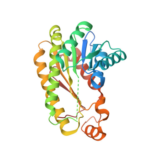
4BNY

Crystal structure of 3-oxoacyl-(acyl-carrier-protein) reductase (FabG) from Pseudomonas aeruginosa in complex with 4-(2-phenylthieno(3,2-d) pyrimidin-4-yl)morpholine at 1.8A resolution


Experimental Data Snapshot

  • Method: X-RAY DIFFRACTION
  • Resolution: 1.80 Å
  • R-Value Free: 0.201 
  • R-Value Work: 0.173 
  • R-Value Observed: 0.174 

Starting Model: experimental
View more details

wwPDB Validation   3D Report Full Report


Ligand Structure Quality Assessment 


This is version 1.2 of the entry. See complete history


Literature

Discovery of an Allosteric Inhibitor Binding Site in 3-Oxo-Acyl-Acp Reductase from Pseudomonas Aeruginosa

Cukier, C.D.Hope, A.Elamin, A.Moynie, L.Schnell, R.Schach, S.Kneuper, H.Singh, M.Naismith, J.Lindqvist, Y.Gray, D.Schneider, G.

(2013) ACS Chem Biol 8: 2518

  • DOI: https://doi.org/10.1021/cb4005063
  • Primary Citation of Related Structures:  
    4AFN, 4AG3, 4BNT, 4BNU, 4BNV, 4BNW, 4BNX, 4BNY, 4BNZ, 4BO0, 4BO1, 4BO2, 4BO3, 4BO4, 4BO5, 4BO6, 4BO7, 4BO8, 4BO9

  • PubMed Abstract: 

    3-Oxo-acyl-acyl carrier protein (ACP) reductase (FabG) plays a key role in the bacterial fatty acid synthesis II system in pathogenic microorganisms, which has been recognized as a potential drug target. FabG catalyzes reduction of a 3-oxo-acyl-ACP intermediate during the elongation cycle of fatty acid biosynthesis. Here, we report gene deletion experiments that support the essentiality of this gene in P. aeruginosa and the identification of a number of small molecule FabG inhibitors with IC50 values in the nanomolar to low micromolar range and good physicochemical properties. Structural characterization of 16 FabG-inhibitor complexes by X-ray crystallography revealed that the compounds bind at a novel allosteric site located at the FabG subunit-subunit interface. Inhibitor binding relies primarily on hydrophobic interactions, but specific hydrogen bonds are also observed. Importantly, the binding cavity is formed upon complex formation and therefore would not be recognized by virtual screening approaches. The structure analysis further reveals that the inhibitors act by inducing conformational changes that propagate to the active site, resulting in a displacement of the catalytic triad and the inability to bind NADPH.


  • Organizational Affiliation

    Department of Medical Biochemistry and Biophysics, Karolinska Institutet , 17177 Stockholm, Sweden.


Macromolecules
Find similar proteins by:  (by identity cutoff)  |  3D Structure
Entity ID: 1
MoleculeChains Sequence LengthOrganismDetailsImage
3-OXOACYL-[ACYL-CARRIER-PROTEIN] REDUCTASE FABG
A, B, C, D
269Pseudomonas aeruginosa PAO1Mutation(s): 0 
EC: 1.1.1.100
UniProt
Find proteins for O54438 (Pseudomonas aeruginosa (strain ATCC 15692 / DSM 22644 / CIP 104116 / JCM 14847 / LMG 12228 / 1C / PRS 101 / PAO1))
Explore O54438 
Go to UniProtKB:  O54438
Entity Groups  
Sequence Clusters30% Identity50% Identity70% Identity90% Identity95% Identity100% Identity
UniProt GroupO54438
Sequence Annotations
Expand
  • Reference Sequence
Small Molecules
Ligands 1 Unique
IDChains Name / Formula / InChI Key2D Diagram3D Interactions
36I
Query on 36I

Download Ideal Coordinates CCD File 
E [auth B],
F [auth D]
4-(2-phenylthieno[3,2-d]pyrimidin-4-yl)morpholine
C16 H15 N3 O S
RBDRSWPTRGNKIG-UHFFFAOYSA-N
Binding Affinity Annotations 
IDSourceBinding Affinity
36I PDBBind:  4BNY Ki: 80 (nM) from 1 assay(s)
Experimental Data & Validation

Experimental Data

  • Method: X-RAY DIFFRACTION
  • Resolution: 1.80 Å
  • R-Value Free: 0.201 
  • R-Value Work: 0.173 
  • R-Value Observed: 0.174 
  • Space Group: P 21 21 21
Unit Cell:
Length ( Å )Angle ( ˚ )
a = 54.94α = 90
b = 108.39β = 90
c = 148.17γ = 90
Software Package:
Software NamePurpose
REFMACrefinement
iMOSFLMdata reduction
SCALAdata scaling
PHASERphasing

Structure Validation

View Full Validation Report


Ligand Structure Quality Assessment 


Entry History 

Deposition Data

Revision History  (Full details and data files)

  • Version 1.0: 2013-09-18
    Type: Initial release
  • Version 1.1: 2013-11-27
    Changes: Database references
  • Version 1.2: 2023-12-20
    Changes: Data collection, Database references, Derived calculations, Other, Refinement description