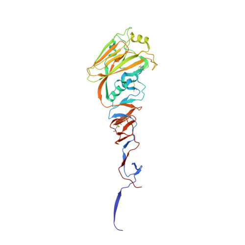
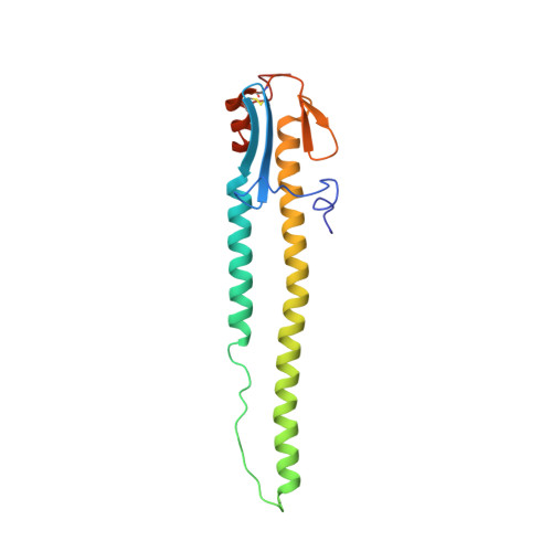
4CQX

H5 (tyTy) Del133/Ile155Thr Mutant Haemagglutinin in Complex with Human Receptor Analogue 6'SLN


Experimental Data Snapshot

  • Method: X-RAY DIFFRACTION
  • Resolution: 2.30 Å
  • R-Value Free: 0.226 
  • R-Value Work: 0.186 
  • R-Value Observed: 0.188 

Starting Model: experimental
View more details

wwPDB Validation   3D Report Full Report


Ligand Structure Quality Assessment 


This is version 2.2 of the entry. See complete history


Literature

Enhanced Human Receptor Binding by H5 Haemagglutinins.

Xiong, X.Xiao, H.Martin, S.R.Coombs, P.J.Liu, J.Collins, P.J.Vachieri, S.G.Walker, P.A.Lin, Y.P.Mccauley, J.W.Gamblin, S.J.Skehel, J.J.

(2014) Virology 456: 179

  • DOI: https://doi.org/10.1016/j.virol.2014.03.008
  • Primary Citation of Related Structures:  
    4CQP, 4CQQ, 4CQR, 4CQS, 4CQU, 4CQV, 4CQW, 4CQX, 4CQY, 4CQZ, 4CR0, 5AJM

  • PubMed Abstract: 

    Mutant H5N1 influenza viruses have been isolated from humans that have increased human receptor avidity. We have compared the receptor binding properties of these mutants with those of wild-type viruses, and determined the structures of their haemagglutinins in complex with receptor analogues. Mutants from Vietnam bind tighter to human receptor by acquiring basic residues near the receptor binding site. They bind more weakly to avian receptor because they lack specific interactions between Asn-186 and Gln-226. In contrast, a double mutant, Δ133/Ile155Thr, isolated in Egypt has greater avidity for human receptor while retaining wild-type avidity for avian receptor. Despite these increases in human receptor binding, none of the mutants prefers human receptor, unlike aerosol transmissible H5N1 viruses. Nevertheless, mutants with high avidity for both human and avian receptors may be intermediates in the evolution of H5N1 viruses that could infect both humans and poultry.


  • Organizational Affiliation

    MRC National Institute for Medical Research, The Ridgeway, Mill Hill, London NW7 1AA, UK.


Macromolecules
Find similar proteins by:  (by identity cutoff)  |  3D Structure
Entity ID: 1
MoleculeChains Sequence LengthOrganismDetailsImage
HAEMAGGLUTININ HA1
A, C, E
327Influenza A virus (A/turkey/Turkey/1/2005(H5N1))Mutation(s): 1 
UniProt
Find proteins for Q207Z6 (Influenza A virus)
Explore Q207Z6 
Go to UniProtKB:  Q207Z6
Entity Groups  
Sequence Clusters30% Identity50% Identity70% Identity90% Identity95% Identity100% Identity
UniProt GroupQ207Z6
Glycosylation
Glycosylation Sites: 3
Sequence Annotations
Expand
  • Reference Sequence
Find similar proteins by:  (by identity cutoff)  |  3D Structure
Entity ID: 2
MoleculeChains Sequence LengthOrganismDetailsImage
HAEMAGGLUTININ HA2
B, D, F
166Influenza A virus (A/turkey/Turkey/1/2005(H5N1))Mutation(s): 0 
UniProt
Find proteins for Q207Z6 (Influenza A virus)
Explore Q207Z6 
Go to UniProtKB:  Q207Z6
Entity Groups  
Sequence Clusters30% Identity50% Identity70% Identity90% Identity95% Identity100% Identity
UniProt GroupQ207Z6
Sequence Annotations
Expand
  • Reference Sequence
Oligosaccharides

Help

Entity ID: 3
MoleculeChains Length2D Diagram Glycosylation3D Interactions
N-acetyl-alpha-neuraminic acid-(2-6)-beta-D-galactopyranose-(1-4)-2-acetamido-2-deoxy-beta-D-glucopyranose
G, H, I
3N/A
Glycosylation Resources
GlyTouCan:  G73578JC
GlyCosmos:  G73578JC
GlyGen:  G73578JC
Small Molecules
Ligands 2 Unique
IDChains Name / Formula / InChI Key2D Diagram3D Interactions
NAG
Query on NAG

Download Ideal Coordinates CCD File 
J [auth A]
K [auth A]
L [auth A]
O [auth C]
P [auth C]
J [auth A],
K [auth A],
L [auth A],
O [auth C],
P [auth C],
S [auth E],
T [auth E],
U [auth E],
V [auth E]
2-acetamido-2-deoxy-beta-D-glucopyranose
C8 H15 N O6
OVRNDRQMDRJTHS-FMDGEEDCSA-N
PO4
Query on PO4

Download Ideal Coordinates CCD File 
M [auth A],
N [auth A],
Q [auth C],
R [auth C],
W [auth E]
PHOSPHATE ION
O4 P
NBIIXXVUZAFLBC-UHFFFAOYSA-K
Experimental Data & Validation

Experimental Data

  • Method: X-RAY DIFFRACTION
  • Resolution: 2.30 Å
  • R-Value Free: 0.226 
  • R-Value Work: 0.186 
  • R-Value Observed: 0.188 
  • Space Group: P 1 21 1
Unit Cell:
Length ( Å )Angle ( ˚ )
a = 87.824α = 90
b = 117.104β = 92.58
c = 101.275γ = 90
Software Package:
Software NamePurpose
REFMACrefinement
xia2data reduction
SCALAdata scaling
PHASERphasing

Structure Validation

View Full Validation Report


Ligand Structure Quality Assessment 


Entry History 

Revision History  (Full details and data files)

  • Version 1.0: 2014-05-28
    Type: Initial release
  • Version 1.1: 2014-06-11
    Changes: Database references
  • Version 2.0: 2020-07-29
    Type: Remediation
    Reason: Carbohydrate remediation
    Changes: Advisory, Atomic model, Data collection, Derived calculations, Other, Structure summary
  • Version 2.1: 2023-12-20
    Changes: Data collection, Database references, Refinement description, Structure summary
  • Version 2.2: 2024-10-23
    Changes: Structure summary