4CUN

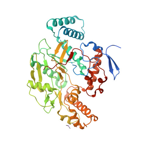
Structure of bovine endothelial nitric oxide synthase heme domain in complex with (9aS)-2-amino-9a-methyl-8,9,9a,10-tetrahydrobenzo[g]pteridine-4,6(3H,7H)-dione


Experimental Data Snapshot

  • Method: X-RAY DIFFRACTION
  • Resolution: 2.48 Å
  • R-Value Free: 0.242 
  • R-Value Work: 0.184 
  • R-Value Observed: 0.187 

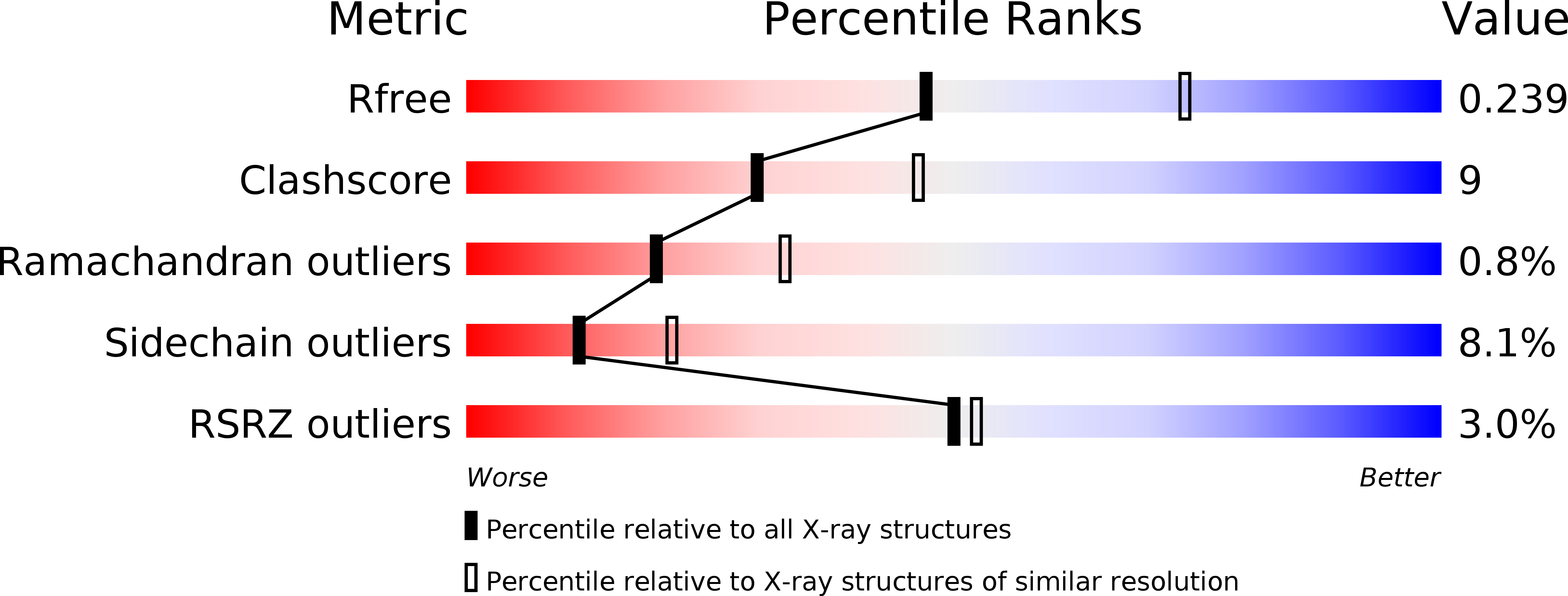
wwPDB Validation   3D Report Full Report


Ligand Structure Quality Assessment 


This is version 1.2 of the entry. See complete history


Literature

Communication between the Zinc and Tetrahydrobiopterin Binding Sites in Nitric Oxide Synthase.

Chreifi, G.Li, H.Mcinnes, C.R.Gibson, C.L.Suckling, C.J.Poulos, T.L.

(2014) Biochemistry 53: 4216

  • DOI: https://doi.org/10.1021/bi5003986
  • Primary Citation of Related Structures:  
    4CUL, 4CUM, 4CUN, 4CVG

  • PubMed Abstract: 

    The nitric oxide synthase (NOS) dimer is stabilized by a Zn(2+) ion coordinated to four symmetry-related Cys residues exactly along the dimer 2-fold axis. Each of the two essential tetrahydrobiopterin (H4B) molecules in the dimer interacts directly with the heme, and each H4B molecule is ~15 Å from the Zn(2+). We have determined the crystal structures of the bovine endothelial NOS dimer oxygenase domain bound to three different pterin analogues, which reveal an intimate structural communication between the H4B and Zn(2+) sites. The binding of one of these compounds, 6-acetyl-2-amino-7,7-dimethyl-7,8-dihydro-4(3H)-pteridinone (1), to the pterin site and Zn(2+) binding are mutually exclusive. Compound 1 both directly and indirectly disrupts hydrogen bonding between key residues in the Zn(2+) binding motif, resulting in destabilization of the dimer and a complete disruption of the Zn(2+) site. Addition of excess Zn(2+) stabilizes the Zn(2+) site at the expense of weakened binding of 1. The unique structural features of 1 that disrupt the dimer interface are extra methyl groups that extend into the dimer interface and force a slight opening of the dimer, thus resulting in disruption of the Zn(2+) site. These results illustrate a very delicate balance of forces and structure at the dimer interface that must be maintained to properly form the Zn(2+), pterin, and substrate binding sites.


  • Organizational Affiliation

    Departments of †Molecular Biology and Biochemistry, ‡Chemistry, and §Pharmaceutical Sciences, University of California , Irvine, California 92697-3900, United States.


Macromolecules
Find similar proteins by:  (by identity cutoff)  |  3D Structure
Entity ID: 1
MoleculeChains Sequence LengthOrganismDetailsImage
NITRIC OXIDE SYNTHASE, ENDOTHELIAL
A, B
443Bos taurusMutation(s): 0 
EC: 1.14.13.39
UniProt
Find proteins for P29473 (Bos taurus)
Explore P29473 
Go to UniProtKB:  P29473
Entity Groups  
Sequence Clusters30% Identity50% Identity70% Identity90% Identity95% Identity100% Identity
UniProt GroupP29473
Sequence Annotations
Expand
  • Reference Sequence
Small Molecules
Ligands 6 Unique
IDChains Name / Formula / InChI Key2D Diagram3D Interactions
HEM
Query on HEM

Download Ideal Coordinates CCD File 
D [auth A],
J [auth B]
PROTOPORPHYRIN IX CONTAINING FE
C34 H32 Fe N4 O4
KABFMIBPWCXCRK-RGGAHWMASA-L
WS6
Query on WS6

Download Ideal Coordinates CCD File 
E [auth A],
K [auth B]
(9aS)-2-amino-9a-methyl-8,9,9a,10-tetrahydrobenzo[g]pteridine-4,6(3H,7H)-dione
C11 H13 N5 O2
FFUUFCSJQNSUEJ-NSHDSACASA-N
ARG
Query on ARG

Download Ideal Coordinates CCD File 
C [auth A],
I [auth B]
ARGININE
C6 H15 N4 O2
ODKSFYDXXFIFQN-BYPYZUCNSA-O
GOL
Query on GOL

Download Ideal Coordinates CCD File 
G [auth A],
M [auth B]
GLYCEROL
C3 H8 O3
PEDCQBHIVMGVHV-UHFFFAOYSA-N
ZN
Query on ZN

Download Ideal Coordinates CCD File 
H [auth A]ZINC ION
Zn
PTFCDOFLOPIGGS-UHFFFAOYSA-N
ACT
Query on ACT

Download Ideal Coordinates CCD File 
F [auth A],
L [auth B]
ACETATE ION
C2 H3 O2
QTBSBXVTEAMEQO-UHFFFAOYSA-M
Modified Residues  1 Unique
IDChains TypeFormula2D DiagramParent
CAS
Query on CAS
A, B
L-PEPTIDE LINKINGC5 H12 As N O2 SCYS
Experimental Data & Validation

Experimental Data

  • Method: X-RAY DIFFRACTION
  • Resolution: 2.48 Å
  • R-Value Free: 0.242 
  • R-Value Work: 0.184 
  • R-Value Observed: 0.187 
  • Space Group: P 21 21 21
Unit Cell:
Length ( Å )Angle ( ˚ )
a = 58.865α = 90
b = 106.177β = 90
c = 156.735γ = 90
Software Package:
Software NamePurpose
REFMACrefinement
HKL-2000data reduction
SCALEPACKdata scaling
REFMACphasing

Structure Validation

View Full Validation Report


Ligand Structure Quality Assessment 


Entry History 

Deposition Data

Revision History  (Full details and data files)

  • Version 1.0: 2014-05-28
    Type: Initial release
  • Version 1.1: 2014-07-16
    Changes: Database references
  • Version 1.2: 2024-11-06
    Changes: Data collection, Database references, Derived calculations, Other, Structure summary