4ETP | pdb_00004etp

C-terminal motor and motor homology domain of Kar3Vik1 fused to a synthetic heterodimeric coiled coil


Experimental Data Snapshot

  • Method: X-RAY DIFFRACTION
  • Resolution: 2.30 Å
  • R-Value Free: 
    0.245 (Depositor), 0.250 (DCC) 
  • R-Value Work: 
    0.207 (Depositor), 0.210 (DCC) 
  • R-Value Observed: 
    0.209 (Depositor) 

Starting Models: experimental
View more details

wwPDB Validation   3D Report Full Report


Ligand Structure Quality Assessment 

Created with Raphaël 2.3.0Worse 01 BetterLigand structure goodness of fit to experimental dataBest fitted ADPClick on this verticalbar to view detailsBest fitted EBCClick on this verticalbar to view details

This is version 1.3 of the entry. See complete history


Literature

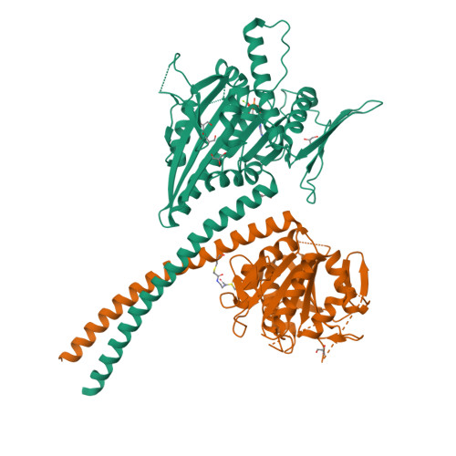
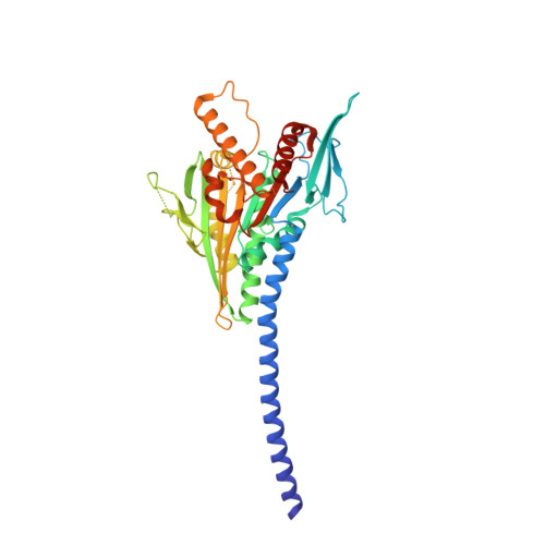
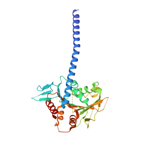
Kar3Vik1, a member of the kinesin-14 superfamily, shows a novel kinesin microtubule binding pattern.

Rank, K.C.Chen, C.J.Cope, J.Porche, K.Hoenger, A.Gilbert, S.P.Rayment, I.

(2012) J Cell Biol 197: 957-970

  • DOI: https://doi.org/10.1083/jcb.201201132
  • Primary Citation of Related Structures:  
    4ETP

  • PubMed Abstract: 

    Kinesin-14 motors generate microtubule minus-end-directed force used in mitosis and meiosis. These motors are dimeric and operate with a nonprocessive powerstroke mechanism, but the role of the second head in motility has been unclear. In Saccharomyces cerevisiae, the Kinesin-14 Kar3 forms a heterodimer with either Vik1 or Cik1. Vik1 contains a motor homology domain that retains microtubule binding properties but lacks a nucleotide binding site. In this case, both heads are implicated in motility. Here, we show through structural determination of a C-terminal heterodimeric Kar3Vik1, electron microscopy, equilibrium binding, and motility that at the start of the cycle, Kar3Vik1 binds to or occludes two αβ-tubulin subunits on adjacent protofilaments. The cycle begins as Vik1 collides with the microtubule followed by Kar3 microtubule association and ADP release, thereby destabilizing the Vik1-microtubule interaction and positioning the motor for the start of the powerstroke. The results indicate that head-head communication is mediated through the adjoining coiled coil.


  • Organizational Affiliation

    Department of Biochemistry, University of Wisconsin, Madison, WI 53706, USA.


Macromolecules
Find similar proteins by:  (by identity cutoff)  |  3D Structure
Entity ID: 1
MoleculeChains Sequence LengthOrganismDetailsImage
Kinesin-like protein KAR3403Saccharomyces cerevisiae S288CMutation(s): 5 
Gene Names: KAR3kar3pP9659.16YPR141C
EC: 5.6.1.4
UniProt
Find proteins for P17119 (Saccharomyces cerevisiae (strain ATCC 204508 / S288c))
Explore P17119 
Go to UniProtKB:  P17119
Entity Groups  
Sequence Clusters30% Identity50% Identity70% Identity90% Identity95% Identity100% Identity
UniProt GroupP17119
Sequence Annotations
Expand
  • Reference Sequence
Find similar proteins by:  (by identity cutoff)  |  3D Structure
Entity ID: 2
MoleculeChains Sequence LengthOrganismDetailsImage
Spindle pole body-associated protein VIK1333Saccharomyces cerevisiae S288CMutation(s): 7 
Gene Names: VIK1Vik1pYPL253C
UniProt
Find proteins for Q12045 (Saccharomyces cerevisiae (strain ATCC 204508 / S288c))
Explore Q12045 
Go to UniProtKB:  Q12045
Entity Groups  
Sequence Clusters30% Identity50% Identity70% Identity90% Identity95% Identity100% Identity
UniProt GroupQ12045
Sequence Annotations
Expand
  • Reference Sequence
Small Molecules
Ligands 4 Unique
IDChains Name / Formula / InChI Key2D Diagram3D Interactions
ADP
Query on ADP

Download Ideal Coordinates CCD File 
C [auth A]ADENOSINE-5'-DIPHOSPHATE
C10 H15 N5 O10 P2
XTWYTFMLZFPYCI-KQYNXXCUSA-N
EBC
Query on EBC

Download Ideal Coordinates CCD File 
J [auth B]N,N'-ethane-1,2-diylbis(2-iodoacetamide)
C6 H10 I2 N2 O2
RLFPCLMBTQOMLI-UHFFFAOYSA-N
GOL
Query on GOL

Download Ideal Coordinates CCD File 
E [auth A]
F [auth A]
G [auth A]
H [auth A]
I [auth A]
GLYCEROL
C3 H8 O3
PEDCQBHIVMGVHV-UHFFFAOYSA-N
MG
Query on MG

Download Ideal Coordinates CCD File 
D [auth A]MAGNESIUM ION
Mg
JLVVSXFLKOJNIY-UHFFFAOYSA-N
Experimental Data & Validation

Experimental Data

  • Method: X-RAY DIFFRACTION
  • Resolution: 2.30 Å
  • R-Value Free:  0.245 (Depositor), 0.250 (DCC) 
  • R-Value Work:  0.207 (Depositor), 0.210 (DCC) 
  • R-Value Observed: 0.209 (Depositor) 
Space Group: P 21 21 21
Unit Cell:
Length ( Å )Angle ( ˚ )
a = 88.06α = 90
b = 94.939β = 90
c = 114.59γ = 90
Software Package:
Software NamePurpose
HKL-2000data collection
PHASERphasing
REFMACrefinement
HKL-2000data reduction
HKL-2000data scaling

Structure Validation

View Full Validation Report


Ligand Structure Quality Assessment 

Created with Raphaël 2.3.0Worse 01 BetterLigand structure goodness of fit to experimental dataBest fitted ADPClick on this verticalbar to view detailsBest fitted EBCClick on this verticalbar to view details

Entry History 

Deposition Data

Revision History  (Full details and data files)

  • Version 1.0: 2012-06-27
    Type: Initial release
  • Version 1.1: 2013-08-07
    Changes: Database references
  • Version 1.2: 2023-09-13
    Changes: Data collection, Database references, Derived calculations, Refinement description
  • Version 1.3: 2024-11-20
    Changes: Structure summary