4I3B | pdb_00004i3b

Crystal structure of fluorescent protein UnaG wild type


Experimental Data Snapshot

  • Method: X-RAY DIFFRACTION
  • Resolution: 1.20 Å
  • R-Value Free: 
    0.159 (Depositor), 0.160 (DCC) 
  • R-Value Work: 
    0.129 (Depositor), 0.130 (DCC) 
  • R-Value Observed: 
    0.131 (Depositor) 

Starting Model: experimental
View more details

wwPDB Validation   3D Report Full Report


Ligand Structure Quality Assessment 

Created with Raphaël 2.3.0Worse 01 BetterLigand structure goodness of fit to experimental dataBest fitted BLRClick on this verticalbar to view details

This is version 1.3 of the entry. See complete history


Literature

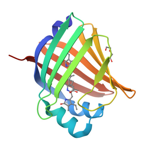
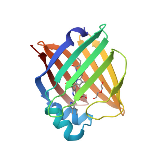
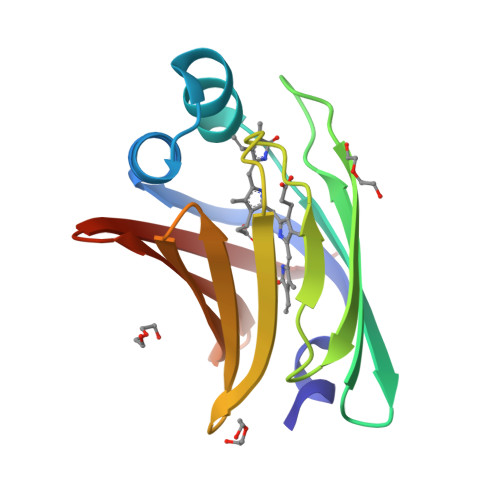
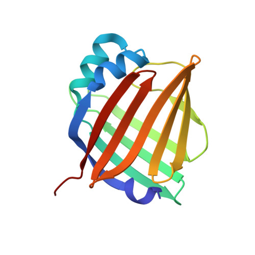
A bilirubin-inducible fluorescent protein from eel muscle

Kumagai, A.Ando, R.Miyatake, H.Greimel, P.Kobayashi, T.Hirabayashi, Y.Shimogori, T.Miyawaki, A.

(2013) Cell 153: 1602-1611

  • DOI: https://doi.org/10.1016/j.cell.2013.05.038
  • Primary Citation of Related Structures:  
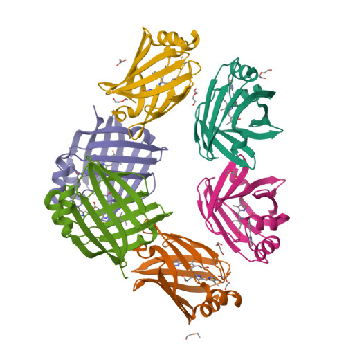
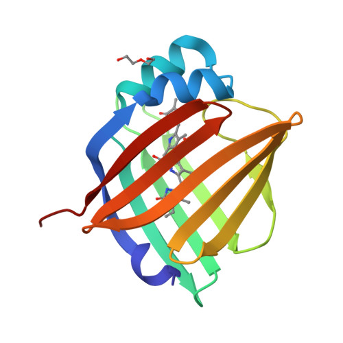
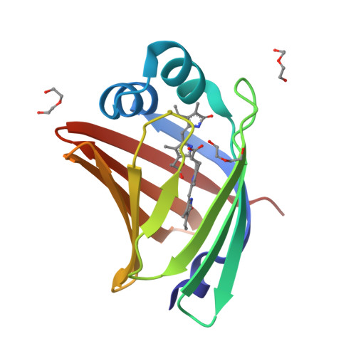
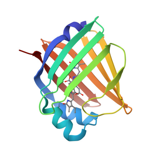
    4I3B, 4I3C, 4I3D

  • PubMed Abstract: 

    The fluorescent protein toolbox has revolutionized experimental biology. Despite this advance, no fluorescent proteins have been identified from vertebrates, nor has chromogenic ligand-inducible activation or clinical utility been demonstrated. Here, we report the cloning and characterization of UnaG, a fluorescent protein from Japanese eel. UnaG belongs to the fatty-acid-binding protein (FABP) family, and expression in eel is restricted to small-diameter muscle fibers. On heterologous expression in cell lines or mouse brain, UnaG produces oxygen-independent green fluorescence. Remarkably, UnaG fluorescence is triggered by an endogenous ligand, bilirubin, a membrane-permeable heme metabolite and clinical health biomarker. The holoUnaG structure at 1.2 Å revealed a biplanar coordination of bilirubin by reversible π-conjugation, and we used this high-affinity and high-specificity interaction to establish a fluorescence-based human bilirubin assay with promising clinical utility. UnaG will be the prototype for a versatile class of ligand-activated fluorescent proteins, with applications in research, medicine, and bioengineering.


  • Organizational Affiliation

    Cell Function Dynamics, Brain Science Institute, RIKEN, 2-1 Hirosawa, Wako-city, Saitama 351-0198, Japan.


Macromolecules
Find similar proteins by:  (by identity cutoff)  |  3D Structure
Entity ID: 1
MoleculeChains Sequence LengthOrganismDetailsImage
Bilirubin-inducible fluorescent protein UnaG
A, B, C, D, E
139Anguilla japonicaMutation(s): 0 
UniProt
Find proteins for P0DM59 (Anguilla japonica)
Explore P0DM59 
Go to UniProtKB:  P0DM59
Entity Groups  
Sequence Clusters30% Identity50% Identity70% Identity90% Identity95% Identity100% Identity
UniProt GroupP0DM59
Sequence Annotations
Expand
  • Reference Sequence
Small Molecules
Ligands 2 Unique
IDChains Name / Formula / InChI Key2D Diagram3D Interactions
BLR
Query on BLR

Download Ideal Coordinates CCD File 
G [auth A]
I [auth B]
M [auth C]
N [auth D]
P [auth E]
3-[5-[(Z)-(4-ethenyl-3-methyl-5-oxidanylidene-pyrrol-2-ylidene)methyl]-2-[[5-[(Z)-(3-ethenyl-4-methyl-5-oxidanylidene-pyrrol-2-ylidene)methyl]-3-(3-hydroxy-3-oxopropyl)-4-methyl-1H-pyrrol-2-yl]methyl]-4-methyl-1H-pyrrol-3-yl]propanoic acid
C33 H36 N4 O6
BPYKTIZUTYGOLE-IFADSCNNSA-N
PEG
Query on PEG

Download Ideal Coordinates CCD File 
H [auth A]
J [auth B]
K [auth B]
L [auth B]
O [auth D]
DI(HYDROXYETHYL)ETHER
C4 H10 O3
MTHSVFCYNBDYFN-UHFFFAOYSA-N
Experimental Data & Validation

Experimental Data

  • Method: X-RAY DIFFRACTION
  • Resolution: 1.20 Å
  • R-Value Free:  0.159 (Depositor), 0.160 (DCC) 
  • R-Value Work:  0.129 (Depositor), 0.130 (DCC) 
  • R-Value Observed: 0.131 (Depositor) 
Space Group: P 1 21 1
Unit Cell:
Length ( Å )Angle ( ˚ )
a = 44.394α = 90
b = 73.954β = 92.45
c = 124.364γ = 90
Software Package:
Software NamePurpose
HKL-2000data collection
PHENIXmodel building
PHENIXrefinement
HKL-2000data reduction
HKL-2000data scaling
PHENIXphasing

Structure Validation

View Full Validation Report


Ligand Structure Quality Assessment 

Created with Raphaël 2.3.0Worse 01 BetterLigand structure goodness of fit to experimental dataBest fitted BLRClick on this verticalbar to view details

Entry History 

Deposition Data

Revision History  (Full details and data files)

  • Version 1.0: 2013-06-19
    Type: Initial release
  • Version 1.1: 2013-07-31
    Changes: Database references
  • Version 1.2: 2015-02-04
    Changes: Database references, Non-polymer description
  • Version 1.3: 2023-11-08
    Changes: Data collection, Database references, Derived calculations, Refinement description