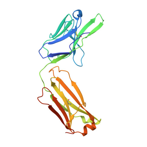
4IMK

Uncrossed Fab binding to human Angiopoietin 2


Experimental Data Snapshot

  • Method: X-RAY DIFFRACTION
  • Resolution: 2.20 Å
  • R-Value Free: 0.226 
  • R-Value Work: 0.199 
  • R-Value Observed: 0.200 

wwPDB Validation   3D Report Full Report


This is version 1.1 of the entry. See complete history


Literature

Crystal Structure of an Anti-Ang2 CrossFab Demonstrates Complete Structural and Functional Integrity of the Variable Domain.

Fenn, S.Schiller, C.B.Griese, J.J.Duerr, H.Imhof-Jung, S.Gassner, C.Moelleken, J.Regula, J.T.Schaefer, W.Thomas, M.Klein, C.Hopfner, K.P.Kettenberger, H.

(2013) PLoS One 8: e61953-e61953

  • DOI: https://doi.org/10.1371/journal.pone.0061953
  • Primary Citation of Related Structures:  
    4IMK, 4IML

  • PubMed Abstract: 

    Bispecific antibodies are considered as a promising class of future biotherapeutic molecules. They comprise binding specificities for two different antigens, which may provide additive or synergistic modes of action. There is a wide variety of design alternatives for such bispecific antibodies, including the "CrossMab" format. CrossMabs contain a domain crossover in one of the antigen-binding (Fab) parts, together with the "knobs-and-holes" approach, to enforce the correct assembly of four different polypeptide chains into an IgG-like bispecific antibody. We determined the crystal structure of a hAng-2-binding Fab in its crossed and uncrossed form and show that CH1-CL-domain crossover does not induce significant perturbations of the structure and has no detectable influence on target binding.


  • Organizational Affiliation

    Large Molecule Research, Pharma Research and Early Development, Roche Dignostics GmbH, Penzberg, Germany.


Macromolecules
Find similar proteins by:  (by identity cutoff)  |  3D Structure
Entity ID: 1
MoleculeChains Sequence LengthOrganismDetailsImage
Heavy Chain
A, B
230Homo sapiensMutation(s): 0 
Entity Groups  
Sequence Clusters30% Identity50% Identity70% Identity90% Identity95% Identity100% Identity
Sequence Annotations
Expand
  • Reference Sequence
Find similar proteins by:  (by identity cutoff)  |  3D Structure
Entity ID: 2
MoleculeChains Sequence LengthOrganismDetailsImage
Light Chain
C, D
224Homo sapiensMutation(s): 0 
Entity Groups  
Sequence Clusters30% Identity50% Identity70% Identity90% Identity95% Identity100% Identity
Sequence Annotations
Expand
  • Reference Sequence
Experimental Data & Validation

Experimental Data

  • Method: X-RAY DIFFRACTION
  • Resolution: 2.20 Å
  • R-Value Free: 0.226 
  • R-Value Work: 0.199 
  • R-Value Observed: 0.200 
  • Space Group: P 21 21 21
Unit Cell:
Length ( Å )Angle ( ˚ )
a = 65.969α = 90
b = 86.67β = 90
c = 205.813γ = 90
Software Package:
Software NamePurpose
PHASERphasing
PHENIXrefinement
XDSdata reduction
XDSdata scaling

Structure Validation

View Full Validation Report


Entry History 

Deposition Data

Revision History  (Full details and data files)

  • Version 1.0: 2013-04-17
    Type: Initial release
  • Version 1.1: 2013-05-22
    Changes: Database references