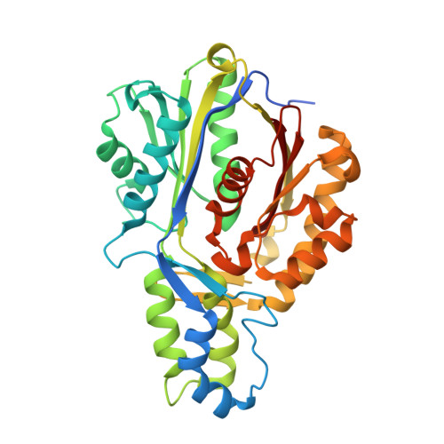
4KU5

Crystal Structures of C143S Xanthomonas campestris OleA with Bound Lauric Acid and Lauroyl-CoA


Experimental Data Snapshot

  • Method: X-RAY DIFFRACTION
  • Resolution: 2.17 Å
  • R-Value Free: 0.203 
  • R-Value Work: 0.159 
  • R-Value Observed: 0.161 

Starting Model: experimental
View more details

wwPDB Validation   3D Report Full Report


Ligand Structure Quality Assessment 


This is version 1.2 of the entry. See complete history


Literature

Substrate Trapping in Crystals of the Thiolase OleA Identifies Three Channels That Enable Long Chain Olefin Biosynthesis.

Goblirsch, B.R.Jensen, M.R.Mohamed, F.A.Wackett, L.P.Wilmot, C.M.

(2016) J Biol Chem 291: 26698-26706

  • DOI: https://doi.org/10.1074/jbc.M116.760892
  • Primary Citation of Related Structures:  
    4KTI, 4KTM, 4KU2, 4KU3, 4KU5

  • PubMed Abstract: 

    Phylogenetically diverse microbes that produce long chain, olefinic hydrocarbons have received much attention as possible sources of renewable energy biocatalysts. One enzyme that is critical for this process is OleA, a thiolase superfamily enzyme that condenses two fatty acyl-CoA substrates to produce a β-ketoacid product and initiates the biosynthesis of long chain olefins in bacteria. Thiolases typically utilize a ping-pong mechanism centered on an active site cysteine residue. Reaction with the first substrate produces a covalent cysteine-thioester tethered acyl group that is transferred to the second substrate through formation of a carbon-carbon bond. Although the basics of thiolase chemistry are precedented, the mechanism by which OleA accommodates two substrates with extended carbon chains and a coenzyme moiety-unusual for a thiolase-are unknown. Gaining insights into this process could enable manipulation of the system for large scale olefin production with hydrocarbon chains lengths equivalent to those of fossil fuels. In this study, mutagenesis of the active site cysteine in Xanthomonas campestris OleA (Cys 143 ) enabled trapping of two catalytically relevant species in crystals. In the resulting structures, long chain alkyl groups (C 12 and C 14 ) and phosphopantetheinate define three substrate channels in a T-shaped configuration, explaining how OleA coordinates its two substrates and product. The C143A OleA co-crystal structure possesses a single bound acyl-CoA representing the Michaelis complex with the first substrate, whereas the C143S co-crystal structure contains both acyl-CoA and fatty acid, defining how a second substrate binds to the acyl-enzyme intermediate. An active site glutamate (Gluβ 117 ) is positioned to deprotonate bound acyl-CoA and initiate carbon-carbon bond formation.


  • Organizational Affiliation

    From the Department of Biochemistry, Molecular Biology, and Biophysics and Biotechnology Institute, University of Minnesota, St. Paul, Minnesota 55108.


Macromolecules
Find similar proteins by:  (by identity cutoff)  |  3D Structure
Entity ID: 1
MoleculeChains Sequence LengthOrganismDetailsImage
3-oxoacyl-[ACP] synthase III
A, B
358Xanthomonas campestris pv. campestris str. ATCC 33913Mutation(s): 1 
Gene Names: fabHXCC0212
EC: 2.3.3.20
UniProt
Find proteins for Q8PDX2 (Xanthomonas campestris pv. campestris (strain ATCC 33913 / DSM 3586 / NCPPB 528 / LMG 568 / P 25))
Explore Q8PDX2 
Go to UniProtKB:  Q8PDX2
Entity Groups  
Sequence Clusters30% Identity50% Identity70% Identity90% Identity95% Identity100% Identity
UniProt GroupQ8PDX2
Sequence Annotations
Expand
  • Reference Sequence
Small Molecules
Ligands 4 Unique
IDChains Name / Formula / InChI Key2D Diagram3D Interactions
DCC
Query on DCC

Download Ideal Coordinates CCD File 
I [auth B]DODECYL-COA
C33 H58 N7 O17 P3 S
YMCXGHLSVALICC-GMHMEAMDSA-N
DAO
Query on DAO

Download Ideal Coordinates CCD File 
G [auth B]LAURIC ACID
C12 H24 O2
POULHZVOKOAJMA-UHFFFAOYSA-N
PEG
Query on PEG

Download Ideal Coordinates CCD File 
C [auth A],
D [auth A],
E [auth A],
H [auth B],
J [auth B]
DI(HYDROXYETHYL)ETHER
C4 H10 O3
MTHSVFCYNBDYFN-UHFFFAOYSA-N
PO4
Query on PO4

Download Ideal Coordinates CCD File 
F [auth A]PHOSPHATE ION
O4 P
NBIIXXVUZAFLBC-UHFFFAOYSA-K
Experimental Data & Validation

Experimental Data

  • Method: X-RAY DIFFRACTION
  • Resolution: 2.17 Å
  • R-Value Free: 0.203 
  • R-Value Work: 0.159 
  • R-Value Observed: 0.161 
  • Space Group: P 21 21 21
Unit Cell:
Length ( Å )Angle ( ˚ )
a = 82.204α = 90
b = 85.379β = 90
c = 102.706γ = 90
Software Package:
Software NamePurpose
Blu-Icedata collection
PHENIXmodel building
PHENIXrefinement
HKL-2000data reduction
HKL-2000data scaling
PHENIXphasing

Structure Validation

View Full Validation Report


Ligand Structure Quality Assessment 


Entry History 

Deposition Data

Revision History  (Full details and data files)

  • Version 1.0: 2014-07-23
    Type: Initial release
  • Version 1.1: 2017-01-11
    Changes: Database references
  • Version 1.2: 2023-09-20
    Changes: Data collection, Database references, Derived calculations, Refinement description