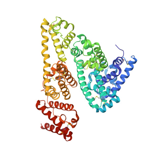
4L9Q

X-ray study of human serum albumin complexed with teniposide


Experimental Data Snapshot

  • Method: X-RAY DIFFRACTION
  • Resolution: 2.70 Å
  • R-Value Free: 0.243 
  • R-Value Work: 0.187 
  • R-Value Observed: 0.190 

Starting Model: experimental
View more details

wwPDB Validation   3D Report Full Report


Ligand Structure Quality Assessment 


This is version 1.4 of the entry. See complete history


Literature

Structural studies of several clinically important oncology drugs in complex with human serum albumin.

Wang, Z.M.Ho, J.X.Ruble, J.R.Rose, J.Ruker, F.Ellenburg, M.Murphy, R.Click, J.Soistman, E.Wilkerson, L.Carter, D.C.

(2013) Biochim Biophys Acta 1830: 5356-5374

  • DOI: https://doi.org/10.1016/j.bbagen.2013.06.032
  • Primary Citation of Related Structures:  
    4L8U, 4L9K, 4L9Q, 4LA0, 4LB2, 4LB9

  • PubMed Abstract: 

    Serum albumin is a major pharmacokinetic effector of drugs. To gain further insight into albumin binding chemistry, the crystal structures of six oncology agents were determined in complex with human serum albumin at resolutions of 2.8 to 2.0Å: camptothecin, 9-amino-camptothecin, etoposide, teniposide, bicalutamide and idarubicin. Protein crystal growth and low temperature X-ray crystallography These large, complex drugs are all bound within the subdomain IB binding region which can be described as a hydrophobic groove formed by α-helices h7, h8 and h9 covered by the extended polypeptide L1. L1 creates a binding cavity with two access sites, one between loop L1 and α-helices h7 and h8 (distal site: IBd) and the other between L1 and α-helix h9 (proximal site: IBp). Camptothecin (2.4Å) and 9 amino camptothecin (2.0Å) are clearly bound as the open lactone form (IBp). Idarubicin (2.8Å) binds in a DNA like dimer complex via an intermolecular π stacking arrangement in IBd. Bicalutamide (2.4Å) is bound in a folded intramolecular π stacking arrangement between two aromatic rings in IBd similar to idarubicin. Teniposide (2.7Å) and etoposide (2.7Å), despite small chemical differences, are bound in two distinctly different sites at or near IB. Teniposide is internalized via primarily hydrophobic interactions and spans through both openings (IBp-d). Etoposide is bound between the exterior of IB and IIA and exhibits an extensive hydrogen bonding network. Subdomain IB is a major binding site for complex heterocyclic molecules. The structures have important implications for drug design and development. This article is part of a Special Issue entitled Serum Albumin.


  • Organizational Affiliation

    New Century Pharmaceuticals, Inc., 895 Martin Road, Suite C, Huntsville, AL 35824, USA.


Macromolecules
Find similar proteins by:  (by identity cutoff)  |  3D Structure
Entity ID: 1
MoleculeChains Sequence LengthOrganismDetailsImage
SERUM ALBUMIN
A, B
585Homo sapiensMutation(s): 0 
UniProt & NIH Common Fund Data Resources
Find proteins for P02768 (Homo sapiens)
Explore P02768 
Go to UniProtKB:  P02768
PHAROS:  P02768
GTEx:  ENSG00000163631 
Entity Groups  
Sequence Clusters30% Identity50% Identity70% Identity90% Identity95% Identity100% Identity
UniProt GroupP02768
Sequence Annotations
Expand
  • Reference Sequence
Small Molecules
Ligands 1 Unique
IDChains Name / Formula / InChI Key2D Diagram3D Interactions
9TP
Query on 9TP

Download Ideal Coordinates CCD File 
C [auth A],
D [auth B]
(5S,5aR,8aR,9R)-9-(4-hydroxy-3,5-dimethoxyphenyl)-8-oxo-5,5a,6,8,8a,9-hexahydrofuro[3',4':6,7]naphtho[2,3-d][1,3]dioxol -5-yl 4,6-O-(thiophen-2-ylmethylidene)-beta-D-glucopyranoside
C32 H32 O13 S
NRUKOCRGYNPUPR-QBPJDGROSA-N
Experimental Data & Validation

Experimental Data

  • Method: X-RAY DIFFRACTION
  • Resolution: 2.70 Å
  • R-Value Free: 0.243 
  • R-Value Work: 0.187 
  • R-Value Observed: 0.190 
  • Space Group: P 1
Unit Cell:
Length ( Å )Angle ( ˚ )
a = 56.353α = 73.17
b = 59.588β = 83.45
c = 95.539γ = 73.93
Software Package:
Software NamePurpose
DENZOdata reduction
SCALEPACKdata scaling
PHENIXrefinement
PDB_EXTRACTdata extraction
CrystalCleardata collection
MERLOTphasing

Structure Validation

View Full Validation Report


Ligand Structure Quality Assessment 


Entry History 

Deposition Data

Revision History  (Full details and data files)

  • Version 1.0: 2013-07-24
    Type: Initial release
  • Version 1.1: 2013-10-23
    Changes: Database references
  • Version 1.2: 2014-02-19
    Changes: Other
  • Version 1.3: 2024-04-03
    Changes: Data collection, Database references, Derived calculations, Refinement description, Structure summary
  • Version 1.4: 2024-10-30
    Changes: Structure summary