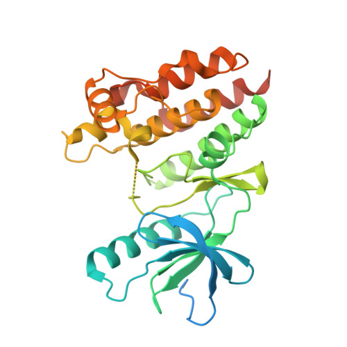
4MNF

Crystal structure of BRAF-V600E bound to GDC0879


Experimental Data Snapshot

  • Method: X-RAY DIFFRACTION
  • Resolution: 2.80 Å
  • R-Value Free: 0.275 
  • R-Value Work: 0.211 
  • R-Value Observed: 0.215 

Starting Model: experimental
View more details

wwPDB Validation   3D Report Full Report


Ligand Structure Quality Assessment 


This is version 1.3 of the entry. See complete history


Literature

Structure of the BRAF-MEK Complex Reveals a Kinase Activity Independent Role for BRAF in MAPK Signaling.

Haling, J.R.Sudhamsu, J.Yen, I.Sideris, S.Sandoval, W.Phung, W.Bravo, B.J.Giannetti, A.M.Peck, A.Masselot, A.Morales, T.Smith, D.Brandhuber, B.J.Hymowitz, S.G.Malek, S.

(2014) Cancer Cell 26: 402-413

  • DOI: https://doi.org/10.1016/j.ccr.2014.07.007
  • Primary Citation of Related Structures:  
    4MNE, 4MNF

  • PubMed Abstract: 

    Numerous oncogenic mutations occur within the BRAF kinase domain (BRAF(KD)). Here we show that stable BRAF-MEK1 complexes are enriched in BRAF(WT) and KRAS mutant (MT) cells but not in BRAF(MT) cells. The crystal structure of the BRAF(KD) in a complex with MEK1 reveals a face-to-face dimer sensitive to MEK1 phosphorylation but insensitive to BRAF dimerization. Structure-guided studies reveal that oncogenic BRAF mutations function by bypassing the requirement for BRAF dimerization for activity or weakening the interaction with MEK1. Finally, we show that conformation-specific BRAF inhibitors can sequester a dormant BRAF-MEK1 complex resulting in pathway inhibition. Taken together, these findings reveal a regulatory role for BRAF in the MAPK pathway independent of its kinase activity but dependent on interaction with MEK.


  • Organizational Affiliation

    Department of Biochemical and Cellular Pharmacology, Genentech Inc., 1 DNA Way, South San Francisco, CA 94080, USA.


Macromolecules
Find similar proteins by:  (by identity cutoff)  |  3D Structure
Entity ID: 1
MoleculeChains Sequence LengthOrganismDetailsImage
Serine/threonine-protein kinase B-raf
A, B
329Homo sapiensMutation(s): 1 
Gene Names: BRAFBRAF1RAFB1
EC: 2.7.11.1
UniProt & NIH Common Fund Data Resources
Find proteins for P15056 (Homo sapiens)
Explore P15056 
Go to UniProtKB:  P15056
PHAROS:  P15056
GTEx:  ENSG00000157764 
Entity Groups  
Sequence Clusters30% Identity50% Identity70% Identity90% Identity95% Identity100% Identity
UniProt GroupP15056
Sequence Annotations
Expand
  • Reference Sequence
Small Molecules
Ligands 2 Unique
IDChains Name / Formula / InChI Key2D Diagram3D Interactions
29L
Query on 29L

Download Ideal Coordinates CCD File 
C [auth A],
D [auth B]
2-{4-[(1E)-1-(hydroxyimino)-2,3-dihydro-1H-inden-5-yl]-3-(pyridin-4-yl)-1H-pyrazol-1-yl}ethanol
C19 H18 N4 O2
DEZZLWQELQORIU-RELWKKBWSA-N
CL
Query on CL

Download Ideal Coordinates CCD File 
E [auth B]CHLORIDE ION
Cl
VEXZGXHMUGYJMC-UHFFFAOYSA-M
Experimental Data & Validation

Experimental Data

  • Method: X-RAY DIFFRACTION
  • Resolution: 2.80 Å
  • R-Value Free: 0.275 
  • R-Value Work: 0.211 
  • R-Value Observed: 0.215 
  • Space Group: P 41 21 2
Unit Cell:
Length ( Å )Angle ( ˚ )
a = 99.74α = 90
b = 99.74β = 90
c = 159.032γ = 90
Software Package:
Software NamePurpose
ADSCdata collection
PHASERphasing
PHENIXrefinement
HKL-2000data reduction
HKL-2000data scaling

Structure Validation

View Full Validation Report


Ligand Structure Quality Assessment 


Entry History 

Deposition Data

Revision History  (Full details and data files)

  • Version 1.0: 2014-06-18
    Type: Initial release
  • Version 1.1: 2014-09-03
    Changes: Database references
  • Version 1.2: 2014-09-24
    Changes: Database references
  • Version 1.3: 2023-09-20
    Changes: Data collection, Database references, Derived calculations, Refinement description