4P02

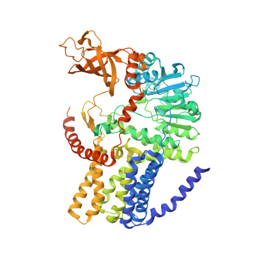
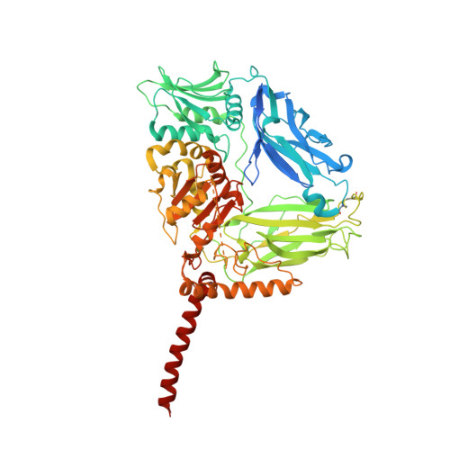
Structure of Bacterial Cellulose Synthase with cyclic-di-GMP bound.


Experimental Data Snapshot

  • Method: X-RAY DIFFRACTION
  • Resolution: 2.65 Å
  • R-Value Free: 0.230 
  • R-Value Work: 0.199 
  • R-Value Observed: 0.200 

wwPDB Validation   3D Report Full Report


Ligand Structure Quality Assessment 


This is version 3.2 of the entry. See complete history


Literature

Mechanism of activation of bacterial cellulose synthase by cyclic di-GMP.

Morgan, J.L.McNamara, J.T.Zimmer, J.

(2014) Nat Struct Mol Biol 21: 489-496

  • DOI: https://doi.org/10.1038/nsmb.2803
  • Primary Citation of Related Structures:  
    4P00, 4P02

  • PubMed Abstract: 

    The bacterial signaling molecule cyclic di-GMP (c-di-GMP) stimulates the synthesis of bacterial cellulose, which is frequently found in biofilms. Bacterial cellulose is synthesized and translocated across the inner membrane by a complex of cellulose synthase BcsA and BcsB subunits. Here we present crystal structures of the c-di-GMP-activated BcsA-BcsB complex. The structures reveal that c-di-GMP releases an autoinhibited state of the enzyme by breaking a salt bridge that otherwise tethers a conserved gating loop that controls access to and substrate coordination at the active site. Disrupting the salt bridge by mutagenesis generates a constitutively active cellulose synthase. Additionally, the c-di-GMP-activated BcsA-BcsB complex contains a nascent cellulose polymer whose terminal glucose unit rests at a new location above BcsA's active site and is positioned for catalysis. Our mechanistic insights indicate how c-di-GMP allosterically modulates enzymatic functions.


  • Organizational Affiliation

    Center for Membrane Biology, Department of Molecular Physiology and Biological Physics, University of Virginia, Charlottesville, Virginia, USA.


Macromolecules
Find similar proteins by:  (by identity cutoff)  |  3D Structure
Entity ID: 1
MoleculeChains Sequence LengthOrganismDetailsImage
Cellulose Synthase subunit A803Cereibacter sphaeroides 2.4.1Mutation(s): 0 
Gene Names: RSP_0333
EC: 2.4.1.12
Membrane Entity: Yes 
UniProt
Find proteins for Q3J125 (Cereibacter sphaeroides (strain ATCC 17023 / DSM 158 / JCM 6121 / CCUG 31486 / LMG 2827 / NBRC 12203 / NCIMB 8253 / ATH 2.4.1.))
Explore Q3J125 
Go to UniProtKB:  Q3J125
Entity Groups  
Sequence Clusters30% Identity50% Identity70% Identity90% Identity95% Identity100% Identity
UniProt GroupQ3J125
Sequence Annotations
Expand
  • Reference Sequence
Find similar proteins by:  (by identity cutoff)  |  3D Structure
Entity ID: 2
MoleculeChains Sequence LengthOrganismDetailsImage
Cellulose Synthase subunit B724Cereibacter sphaeroides 2.4.1Mutation(s): 0 
Gene Names: RSP_0332
Membrane Entity: Yes 
UniProt
Find proteins for Q3J126 (Cereibacter sphaeroides (strain ATCC 17023 / DSM 158 / JCM 6121 / CCUG 31486 / LMG 2827 / NBRC 12203 / NCIMB 8253 / ATH 2.4.1.))
Explore Q3J126 
Go to UniProtKB:  Q3J126
Entity Groups  
Sequence Clusters30% Identity50% Identity70% Identity90% Identity95% Identity100% Identity
UniProt GroupQ3J126
Sequence Annotations
Expand
  • Reference Sequence

Find similar proteins by:  Sequence   |   3D Structure  

Entity ID: 3
MoleculeChains Sequence LengthOrganismDetailsImage
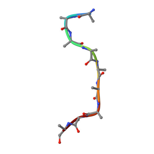
unidentified peptideC [auth D]9Cereibacter sphaeroides 2.4.1Mutation(s): 0 
Sequence Annotations
Expand
  • Reference Sequence
Oligosaccharides

Help

Entity ID: 4
MoleculeChains Length2D Diagram Glycosylation3D Interactions
beta-D-glucopyranose-(1-4)-beta-D-glucopyranose-(1-4)-beta-D-glucopyranose-(1-4)-beta-D-glucopyranose-(1-4)-beta-D-glucopyranose-(1-4)-beta-D-glucopyranose-(1-4)-beta-D-glucopyranose-(1-4)-beta-D-glucopyranose-(1-4)-beta-D-glucopyranose-(1-4)-beta-D-glucopyranose-(1-4)-beta-D-glucopyranose-(1-4)-beta-D-glucopyranose-(1-4)-beta-D-glucopyranose-(1-4)-beta-D-glucopyranose-(1-4)-beta-D-glucopyranose-(1-4)-beta-D-glucopyranose-(1-4)-beta-D-glucopyranoseD [auth C]17N/A
Glycosylation Resources
GlyTouCan:  G14163JN
GlyCosmos:  G14163JN
Small Molecules
Ligands 4 Unique
IDChains Name / Formula / InChI Key2D Diagram3D Interactions
3PE
Query on 3PE

Download Ideal Coordinates CCD File 
H [auth A]1,2-Distearoyl-sn-glycerophosphoethanolamine
C41 H82 N O8 P
LVNGJLRDBYCPGB-LDLOPFEMSA-N
C2E
Query on C2E

Download Ideal Coordinates CCD File 
E [auth A],
F [auth A]
9,9'-[(2R,3R,3aS,5S,7aR,9R,10R,10aS,12S,14aR)-3,5,10,12-tetrahydroxy-5,12-dioxidooctahydro-2H,7H-difuro[3,2-d:3',2'-j][1,3,7,9,2,8]tetraoxadiphosphacyclododecine-2,9-diyl]bis(2-amino-1,9-dihydro-6H-purin-6-one)
C20 H24 N10 O14 P2
PKFDLKSEZWEFGL-MHARETSRSA-N
PLC
Query on PLC

Download Ideal Coordinates CCD File 
G [auth A],
J [auth B],
K [auth B],
L [auth B]
DIUNDECYL PHOSPHATIDYL CHOLINE
C32 H65 N O8 P
IJFVSSZAOYLHEE-SSEXGKCCSA-O
MG
Query on MG

Download Ideal Coordinates CCD File 
I [auth B]MAGNESIUM ION
Mg
JLVVSXFLKOJNIY-UHFFFAOYSA-N
Experimental Data & Validation

Experimental Data

  • Method: X-RAY DIFFRACTION
  • Resolution: 2.65 Å
  • R-Value Free: 0.230 
  • R-Value Work: 0.199 
  • R-Value Observed: 0.200 
  • Space Group: P 21 21 21
Unit Cell:
Length ( Å )Angle ( ˚ )
a = 67.64α = 90
b = 214.66β = 90
c = 220.4γ = 90
Software Package:
Software NamePurpose
PHENIXrefinement

Structure Validation

View Full Validation Report


Ligand Structure Quality Assessment 


Entry History & Funding Information

Deposition Data


Funding OrganizationLocationGrant Number
National Institutes of Health/National Institute of General Medical Sciences (NIH/NIGMS)United StatesR01GM101001
National Science Foundation (NSF, United States)United StatesDGE-1315231.

Revision History  (Full details and data files)

  • Version 1.0: 2014-04-09
    Type: Initial release
  • Version 1.1: 2014-04-23
    Changes: Database references
  • Version 1.2: 2014-07-16
    Changes: Database references
  • Version 1.3: 2014-12-24
    Changes: Database references
  • Version 2.0: 2017-09-27
    Changes: Advisory, Atomic model, Author supporting evidence, Derived calculations, Other, Source and taxonomy, Structure summary
  • Version 2.1: 2019-11-27
    Changes: Author supporting evidence, Data collection
  • Version 3.0: 2020-07-29
    Type: Remediation
    Reason: Carbohydrate remediation
    Changes: Advisory, Atomic model, Data collection, Derived calculations, Refinement description, Structure summary
  • Version 3.1: 2023-12-27
    Changes: Data collection, Database references, Derived calculations, Structure summary
  • Version 3.2: 2024-11-06
    Changes: Structure summary