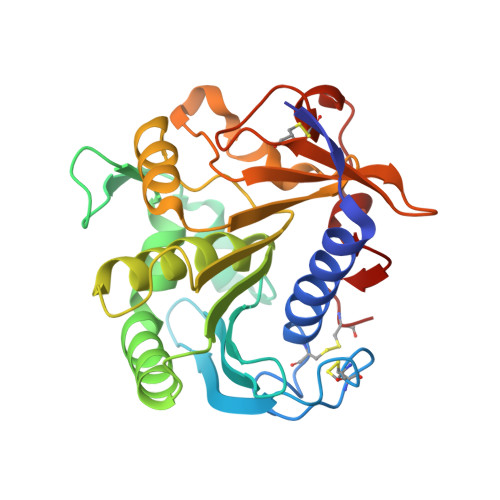
Catalysis at the interface: the anatomy of a conformational change in a triglyceride lipase.
Derewenda, U., Brzozowski, A.M., Lawson, D.M., Derewenda, Z.S.(1992) Biochemistry 31: 1532-1541
- PubMed: 1737010
- DOI: https://doi.org/10.1021/bi00120a034
- Primary Citation of Related Structures:
4TGL - PubMed Abstract:
The crystal structure of an extracellular triglyceride lipase (from a fungus Rhizomucor miehei) inhibited irreversibly by diethyl p-nitrophenyl phosphate (E600) was solved by X-ray crystallographic methods and refined to a resolution of 2.65 A. The crystals are isomorphous with those of n-hexylphosphonate ethyl ester/lipase complex [Brzozowski, A. M., Derewenda, U., Derewenda, Z. S., Dodson, G. G., Lawson, D. M., Turkenburg, J. P., Bjorkling, F., Huge-Jensen, B., Patkar, S. A., & Thim, L. (1991) Nature 351, 491-494], where the conformational change was originally observed. The higher resolution of the present study allowed for a detailed analysis of the stereochemistry of the change observed in the inhibited enzyme. The movement of a 15 amino acid long "lid" (residues 82-96) is a hinge-type rigid-body motion which transports some of the atoms of a short alpha-helix (residues 85-91) by over 12 A. There are two hinge regions (residues 83-84 and 91-95) within which pronounced transitions of secondary structure between alpha and beta conformations are caused by dramatic changes of specific conformational dihedral angles (phi and psi). As a result of this change a hydrophobic area of ca. 800 A2 (8% of the total molecule surface) becomes exposed. Other triglyceride lipases are also known to have "lids" similar to the one observed in the R. miehei enzyme, and it is possible that the general stereochemistry of lipase activation at the oil-water interfaces inferred from the present X-ray study is likely to apply to the entire family of lipases.
Organizational Affiliation:
Department of Biochemistry, University of Alberta, Edmonton, Canada.


