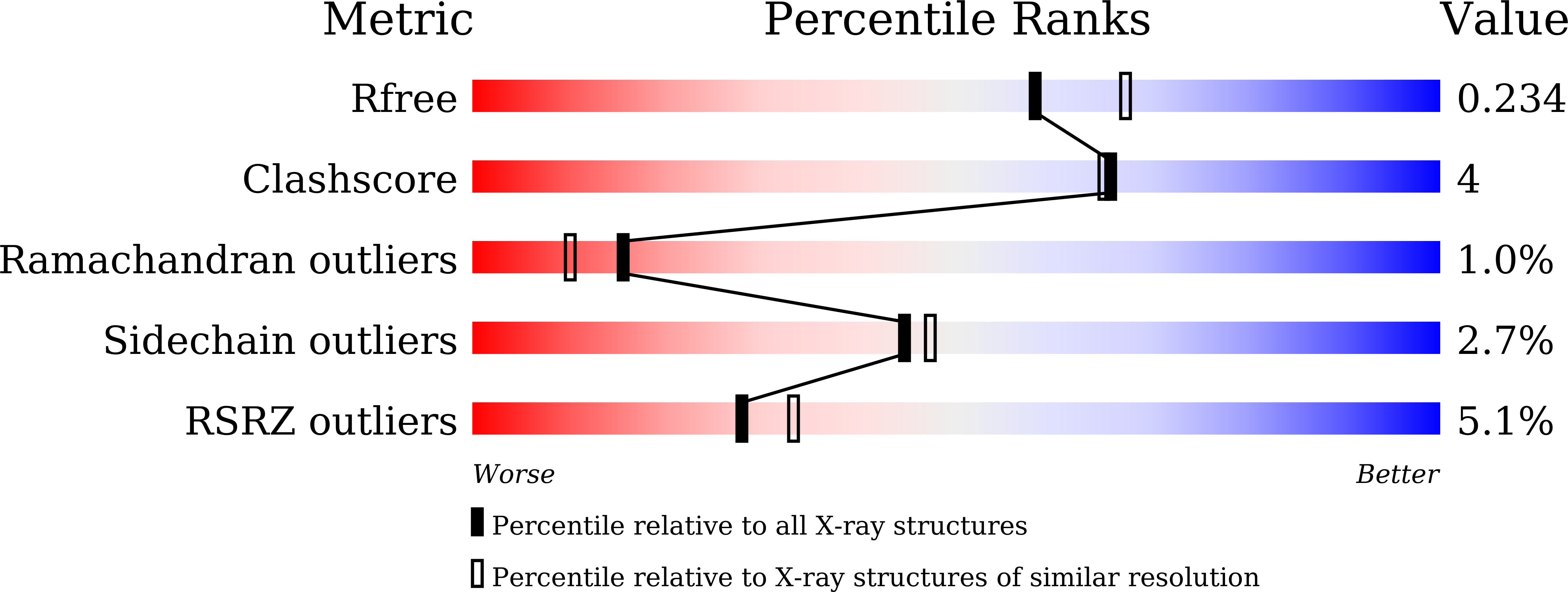
Structures of substrate- and nucleotide-bound propionate kinase from Salmonella typhimurium: substrate specificity and phosphate-transfer mechanism
Murthy, A.M.V., Mathivanan, S., Chittori, S., Savithri, H.S., Murthy, M.R.N.(2015) Acta Crystallogr D Biol Crystallogr 71: 1640-1648
- PubMed: 26249345
- DOI: https://doi.org/10.1107/S1399004715009992
- Primary Citation of Related Structures:
4XH1, 4XH4, 4XH5 - PubMed Abstract:
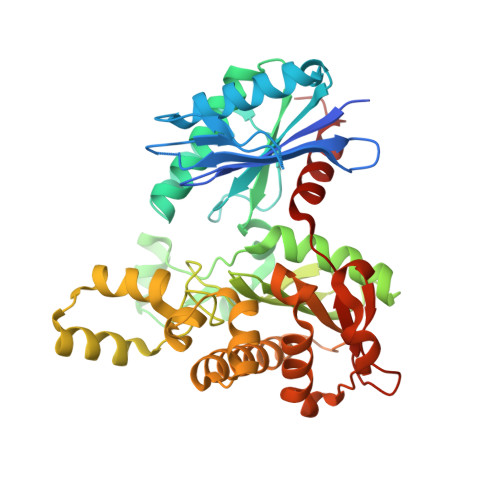
Kinases are ubiquitous enzymes that are pivotal to many biochemical processes. There are contrasting views on the phosphoryl-transfer mechanism in propionate kinase, an enzyme that reversibly transfers a phosphoryl group from propionyl phosphate to ADP in the final step of non-oxidative catabolism of L-threonine to propionate. Here, X-ray crystal structures of propionate- and nucleotide-bound Salmonella typhimurium propionate kinase are reported at 1.8-2.0 Å resolution. Although the mode of nucleotide binding is comparable to those of other members of the ASKHA superfamily, propionate is bound at a distinct site deeper in the hydrophobic pocket defining the active site. The propionate carboxyl is at a distance of ∼ 5 Å from the γ-phosphate of the nucleotide, supporting a direct in-line transfer mechanism. The phosphoryl-transfer reaction is likely to occur via an associative SN2-like transition state that involves a pentagonal bipyramidal structure with the axial positions occupied by the nucleophile of the substrate and the O atom between the β- and the γ-phosphates, respectively. The proximity of the strictly conserved His175 and Arg236 to the carboxyl group of the propionate and the γ-phosphate of ATP suggests their involvement in catalysis. Moreover, ligand binding does not induce global domain movement as reported in some other members of the ASKHA superfamily. Instead, residues Arg86, Asp143 and Pro116-Leu117-His118 that define the active-site pocket move towards the substrate and expel water molecules from the active site. The role of Ala88, previously proposed to be the residue determining substrate specificity, was examined by determining the crystal structures of the propionate-bound Ala88 mutants A88V and A88G. Kinetic analysis and structural data are consistent with a significant role of Ala88 in substrate-specificity determination. The active-site pocket-defining residues Arg86, Asp143 and the Pro116-Leu117-His118 segment are also likely to contribute to substrate specificity.
Organizational Affiliation:
Molecular Biophysics Unit, Indian Institute of Science, Bangalore 560 012, India.


