4ZSI

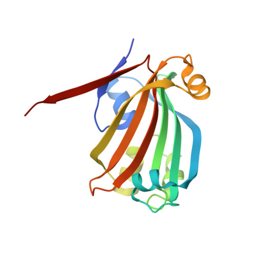
Crystal structure of the effector-binding domain of DasR (DasR-EBD) in complex with glucosamine-6-phosphate


Experimental Data Snapshot

  • Method: X-RAY DIFFRACTION
  • Resolution: 1.65 Å
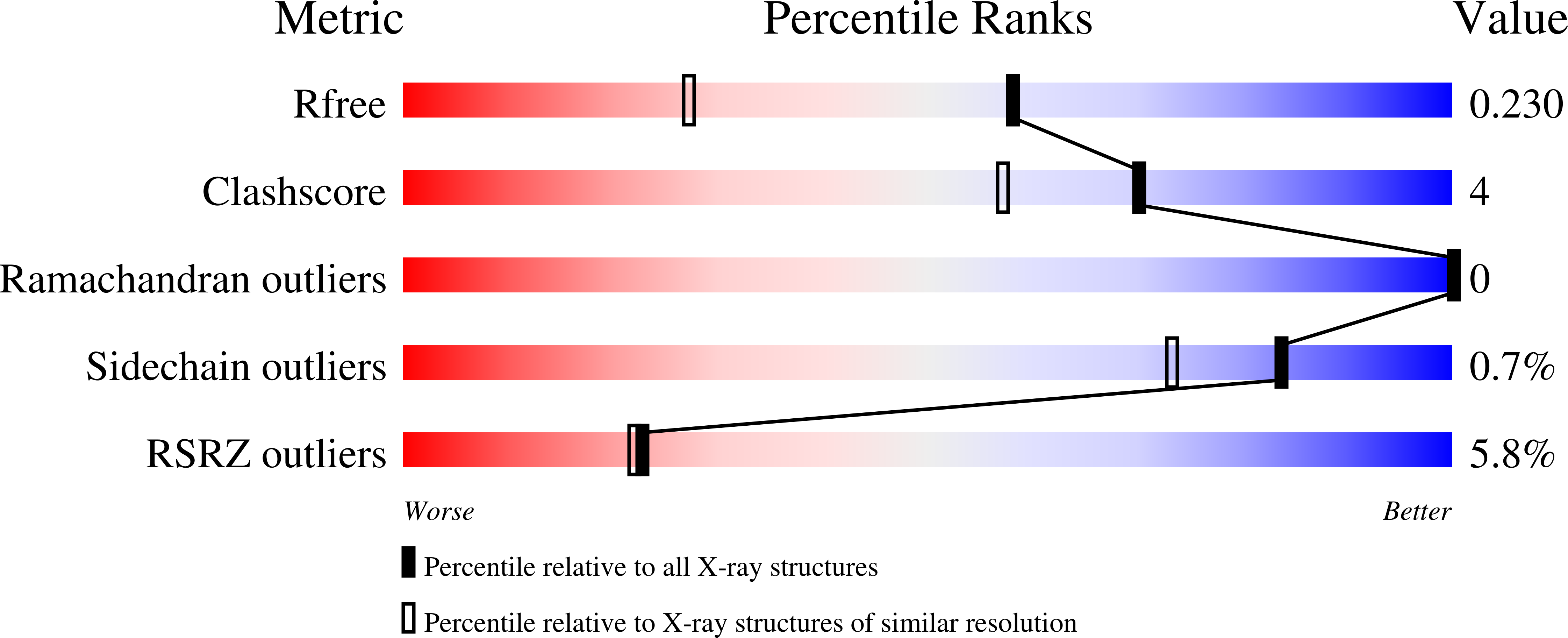
  • R-Value Free: 0.232 
  • R-Value Work: 0.190 
  • R-Value Observed: 0.192 

Starting Model: experimental
View more details

wwPDB Validation   3D Report Full Report


Ligand Structure Quality Assessment 


This is version 1.3 of the entry. See complete history


Literature

Crystal Structures of the Global Regulator DasR from Streptomyces coelicolor: Implications for the Allosteric Regulation of GntR/HutC Repressors.

Fillenberg, S.B.Friess, M.D.Korner, S.Bockmann, R.A.Muller, Y.A.

(2016) PLoS One 11: e0157691-e0157691

  • DOI: https://doi.org/10.1371/journal.pone.0157691
  • Primary Citation of Related Structures:  
    4ZS8, 4ZSB, 4ZSI, 4ZSK

  • PubMed Abstract: 

    Small molecule effectors regulate gene transcription in bacteria by altering the DNA-binding affinities of specific repressor proteins. Although the GntR proteins represent a large family of bacterial repressors, only little is known about the allosteric mechanism that enables their function. DasR from Streptomyces coelicolor belongs to the GntR/HutC subfamily and specifically recognises operators termed DasR-responsive elements (dre-sites). Its DNA-binding properties are modulated by phosphorylated sugars. Here, we present several crystal structures of DasR, namely of dimeric full-length DasR in the absence of any effector and of only the effector-binding domain (EBD) of DasR without effector or in complex with glucosamine-6-phosphate (GlcN-6-P) and N-acetylglucosamine-6-phosphate (GlcNAc-6-P). Together with molecular dynamics (MD) simulations and a comparison with other GntR/HutC family members these data allowed for a structural characterisation of the different functional states of DasR. Allostery in DasR and possibly in many other GntR/HutC family members is best described by a conformational selection model. In ligand-free DasR, an increased flexibility in the EBDs enables the attached DNA-binding domains (DBD) to sample a variety of different orientations and among these also a DNA-binding competent conformation. Effector binding to the EBDs of DasR significantly reorganises the atomic structure of the latter. However, rather than locking the orientation of the DBDs, the effector-induced formation of β-strand β* in the DBD-EBD-linker segment merely appears to take the DBDs 'on a shorter leash' thereby impeding the 'downwards' positioning of the DBDs that is necessary for a concerted binding of two DBDs of DasR to operator DNA.


  • Organizational Affiliation

    Division of Biotechnology, Department of Biology, Friedrich-Alexander University Erlangen-Nuremberg, Henkestr. 91, D-91052 Erlangen, Germany.


Macromolecules
Find similar proteins by:  (by identity cutoff)  |  3D Structure
Entity ID: 1
MoleculeChains Sequence LengthOrganismDetailsImage
HTH-type transcriptional repressor DasR
A, B
171Streptomyces coelicolor A3(2)Mutation(s): 0 
Gene Names: dasRSCO5231SC7E4.28c
UniProt
Find proteins for Q9K492 (Streptomyces coelicolor (strain ATCC BAA-471 / A3(2) / M145))
Explore Q9K492 
Go to UniProtKB:  Q9K492
Entity Groups  
Sequence Clusters30% Identity50% Identity70% Identity90% Identity95% Identity100% Identity
UniProt GroupQ9K492
Sequence Annotations
Expand
  • Reference Sequence
Small Molecules
Ligands 3 Unique
IDChains Name / Formula / InChI Key2D Diagram3D Interactions
4R1
Query on 4R1

Download Ideal Coordinates CCD File 
E [auth B]2-amino-2-deoxy-6-O-phosphono-beta-D-glucopyranose
C6 H14 N O8 P
XHMJOUIAFHJHBW-QZABAPFNSA-N
GLP
Query on GLP

Download Ideal Coordinates CCD File 
C [auth A]2-amino-2-deoxy-6-O-phosphono-alpha-D-glucopyranose
C6 H14 N O8 P
XHMJOUIAFHJHBW-UKFBFLRUSA-N
EDO
Query on EDO

Download Ideal Coordinates CCD File 
D [auth A],
F [auth B]
1,2-ETHANEDIOL
C2 H6 O2
LYCAIKOWRPUZTN-UHFFFAOYSA-N
Experimental Data & Validation

Experimental Data

  • Method: X-RAY DIFFRACTION
  • Resolution: 1.65 Å
  • R-Value Free: 0.232 
  • R-Value Work: 0.190 
  • R-Value Observed: 0.192 
  • Space Group: P 32 2 1
Unit Cell:
Length ( Å )Angle ( ˚ )
a = 54.267α = 90
b = 54.267β = 90
c = 220.913γ = 120
Software Package:
Software NamePurpose
PHENIXrefinement
PHASERphasing
XDSdata reduction
XSCALEdata scaling

Structure Validation

View Full Validation Report


Ligand Structure Quality Assessment 


Entry History & Funding Information

Deposition Data


Funding OrganizationLocationGrant Number
German Research FoundationGermany--

Revision History  (Full details and data files)

  • Version 1.0: 2016-06-08
    Type: Initial release
  • Version 1.1: 2016-07-13
    Changes: Database references
  • Version 1.2: 2020-07-29
    Type: Remediation
    Reason: Carbohydrate remediation
    Changes: Author supporting evidence, Data collection, Derived calculations, Structure summary
  • Version 1.3: 2024-01-10
    Changes: Data collection, Database references, Refinement description, Structure summary