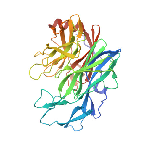
5B0P

Beta-1,2-Mannobiose phosphorylase from Listeria innocua - glycerol complex


Experimental Data Snapshot

  • Method: X-RAY DIFFRACTION
  • Resolution: 1.90 Å
  • R-Value Free: 0.197 
  • R-Value Work: 0.165 
  • R-Value Observed: 0.166 

Starting Model: experimental
View more details

wwPDB Validation   3D Report Full Report


This is version 1.3 of the entry. See complete history


Literature

Characterization and crystal structure determination of beta-1,2-mannobiose phosphorylase from Listeria innocua

Tsuda, T.Nihira, T.Chiku, K.Suzuki, E.Arakawa, T.Nishimoto, M.Kitaoka, M.Nakai, H.Fushinobu, S.

(2015) FEBS Lett 589: 3816-3821

  • DOI: https://doi.org/10.1016/j.febslet.2015.11.034
  • Primary Citation of Related Structures:  
    5B0P, 5B0Q, 5B0R, 5B0S

  • PubMed Abstract: 

    Glycoside hydrolase family 130 consists of phosphorylases and hydrolases for β-mannosides. Here, we characterized β-1,2-mannobiose phosphorylase from Listeria innocua (Lin0857) and determined its crystal structures complexed with β-1,2-linked mannooligosaccharides. β-1,2-Mannotriose was bound in a U-shape, interacting with a phosphate analog at both ends. Lin0857 has a unique dimer structure connected by a loop, and a significant open-close loop displacement was observed for substrate entry. A long loop, which is exclusively present in Lin0857, covers the active site to limit the pocket size. A structural basis for substrate recognition and phosphorolysis was provided.


  • Organizational Affiliation

    Department of Biotechnology, The University of Tokyo, 1-1-1 Yayoi, Bunkyo-ku, Tokyo 113-8657, Japan.


Macromolecules
Find similar proteins by:  (by identity cutoff)  |  3D Structure
Entity ID: 1
MoleculeChains Sequence LengthOrganismDetailsImage
Lin0857 protein
A, B
363Listeria innocua Clip11262Mutation(s): 0 
Gene Names: lin0857
EC: 2.4.1.339
UniProt
Find proteins for Q92DF6 (Listeria innocua serovar 6a (strain ATCC BAA-680 / CLIP 11262))
Explore Q92DF6 
Go to UniProtKB:  Q92DF6
Entity Groups  
Sequence Clusters30% Identity50% Identity70% Identity90% Identity95% Identity100% Identity
UniProt GroupQ92DF6
Sequence Annotations
Expand
  • Reference Sequence
Small Molecules
Ligands 3 Unique
IDChains Name / Formula / InChI Key2D Diagram3D Interactions
MES
Query on MES

Download Ideal Coordinates CCD File 
I [auth A],
J [auth A],
P [auth B],
Q [auth B]
2-(N-MORPHOLINO)-ETHANESULFONIC ACID
C6 H13 N O4 S
SXGZJKUKBWWHRA-UHFFFAOYSA-N
SO4
Query on SO4

Download Ideal Coordinates CCD File 
C [auth A]
D [auth A]
E [auth A]
K [auth B]
L [auth B]
C [auth A],
D [auth A],
E [auth A],
K [auth B],
L [auth B],
M [auth B],
N [auth B]
SULFATE ION
O4 S
QAOWNCQODCNURD-UHFFFAOYSA-L
GOL
Query on GOL

Download Ideal Coordinates CCD File 
F [auth A],
G [auth A],
H [auth A],
O [auth B]
GLYCEROL
C3 H8 O3
PEDCQBHIVMGVHV-UHFFFAOYSA-N
Experimental Data & Validation

Experimental Data

  • Method: X-RAY DIFFRACTION
  • Resolution: 1.90 Å
  • R-Value Free: 0.197 
  • R-Value Work: 0.165 
  • R-Value Observed: 0.166 
  • Space Group: P 31 2 1
Unit Cell:
Length ( Å )Angle ( ˚ )
a = 146.559α = 90
b = 146.559β = 90
c = 106.324γ = 120
Software Package:
Software NamePurpose
REFMACrefinement
HKL-2000data reduction
HKL-2000data scaling
MOLREPphasing

Structure Validation

View Full Validation Report


Entry History & Funding Information

Deposition Data


Funding OrganizationLocationGrant Number
The Ministry of Agriculture, Forestry and Fisheries, JapanJapan25010A

Revision History  (Full details and data files)

  • Version 1.0: 2015-12-02
    Type: Initial release
  • Version 1.1: 2015-12-23
    Changes: Database references
  • Version 1.2: 2020-02-26
    Changes: Data collection, Database references, Derived calculations
  • Version 1.3: 2023-11-08
    Changes: Data collection, Database references, Refinement description