5CFT

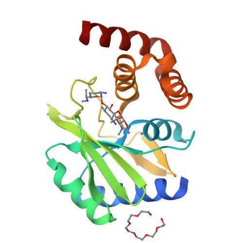
Crystal Structure of ANT(2")-Ia in complex with AMPCPP and gentamicin C1


Experimental Data Snapshot

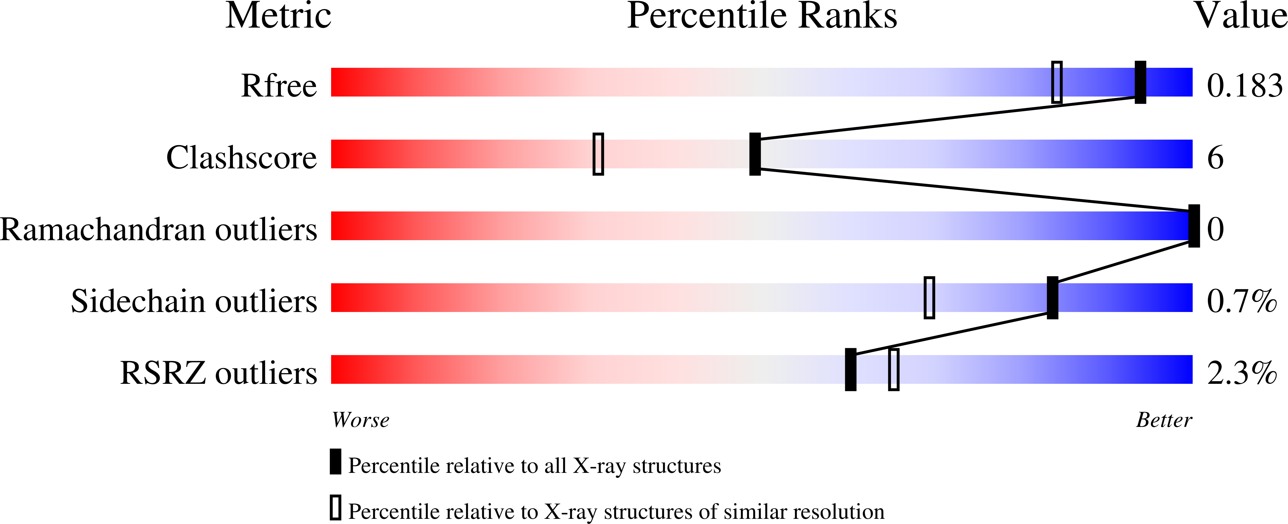
  • Method: X-RAY DIFFRACTION
  • Resolution: 1.50 Å
  • R-Value Free: 0.174 
  • R-Value Work: 0.147 
  • R-Value Observed: 0.149 

wwPDB Validation   3D Report Full Report


Ligand Structure Quality Assessment 


This is version 1.4 of the entry. See complete history


Literature

Structural Analysis of the Tobramycin and Gentamicin Clinical Resistome Reveals Limitations for Next-generation Aminoglycoside Design.

Bassenden, A.V.Rodionov, D.Shi, K.Berghuis, A.M.

(2016) ACS Chem Biol 11: 1339-1346

  • DOI: https://doi.org/10.1021/acschembio.5b01070
  • Primary Citation of Related Structures:  
    5CFS, 5CFT

  • PubMed Abstract: 

    Widespread use and misuse of antibiotics has allowed for the selection of resistant bacteria capable of avoiding the effects of antibiotics. The primary mechanism for resistance to aminoglycosides, a broad-spectrum class of antibiotics, is through covalent enzymatic modification of the drug, waning their bactericidal effect. Tobramycin and gentamicin are two medically important aminoglycosides targeted by several different resistance factors, including aminoglycoside 2″-nucleotidyltransferase [ANT(2″)], the primary cause of aminoglycoside resistance in North America. We describe here two crystal structures of ANT(2″), each in complex with AMPCPP, Mn(2+), and either tobramycin or gentamicin. Together these structures outline ANT(2″)'s specificity for clinically used substrates. Importantly, these structures complete our structural knowledge for the set of enzymes that most frequently confer clinically observed resistance to tobramycin and gentamicin. Comparison of tobramycin and gentamicin binding to enzymes in this resistome, as well as to the intended target, the bacterial ribosome, reveals surprising diversity in observed drug-target interactions. Analysis of the diverse binding modes informs that there are limited opportunities for developing aminoglycoside analogs capable of evading resistance.


  • Organizational Affiliation

    Department of Biochemistry, McGill University , McIntyre Medical Building, 3655 Promenade Sir William Osler, Montreal, Quebec, Canada , H3G 1Y6.


Macromolecules
Find similar proteins by:  (by identity cutoff)  |  3D Structure
Entity ID: 1
MoleculeChains Sequence LengthOrganismDetailsImage
Aminoglycoside Nucleotidyltransferase (2")-Ia185Pseudomonas aeruginosaMutation(s): 0 
Gene Names: aadBTNCP6
UniProt
Find proteins for Q6X3H6 (Pseudomonas aeruginosa)
Explore Q6X3H6 
Go to UniProtKB:  Q6X3H6
Entity Groups  
Sequence Clusters30% Identity50% Identity70% Identity90% Identity95% Identity100% Identity
UniProt GroupQ6X3H6
Sequence Annotations
Expand
  • Reference Sequence
Small Molecules
Ligands 4 Unique
IDChains Name / Formula / InChI Key2D Diagram3D Interactions
APC
Query on APC

Download Ideal Coordinates CCD File 
D [auth A]DIPHOSPHOMETHYLPHOSPHONIC ACID ADENOSYL ESTER
C11 H18 N5 O12 P3
CAWZRIXWFRFUQB-IOSLPCCCSA-N
51G
Query on 51G

Download Ideal Coordinates CCD File 
E [auth A]gentamicin C1
C21 H43 N5 O7
CEAZRRDELHUEMR-CAMVTXANSA-N
P6G
Query on P6G

Download Ideal Coordinates CCD File 
F [auth A]HEXAETHYLENE GLYCOL
C12 H26 O7
IIRDTKBZINWQAW-UHFFFAOYSA-N
MN
Query on MN

Download Ideal Coordinates CCD File 
B [auth A],
C [auth A]
MANGANESE (II) ION
Mn
WAEMQWOKJMHJLA-UHFFFAOYSA-N
Experimental Data & Validation

Experimental Data

  • Method: X-RAY DIFFRACTION
  • Resolution: 1.50 Å
  • R-Value Free: 0.174 
  • R-Value Work: 0.147 
  • R-Value Observed: 0.149 
  • Space Group: P 21 21 21
Unit Cell:
Length ( Å )Angle ( ˚ )
a = 40.58α = 90
b = 46.03β = 90
c = 89γ = 90
Software Package:
Software NamePurpose
PHENIXrefinement
StructureStudiodata collection
xia2data scaling
XSCALEdata scaling
XDSdata reduction
PDB_EXTRACTdata extraction
PHENIXphasing

Structure Validation

View Full Validation Report


Ligand Structure Quality Assessment 


Entry History & Funding Information

Deposition Data


Funding OrganizationLocationGrant Number
Canadian Institutes of Health Research (CIHR)CanadaCIHR MOP-13107

Revision History  (Full details and data files)

  • Version 1.0: 2016-03-16
    Type: Initial release
  • Version 1.1: 2016-06-01
    Changes: Database references
  • Version 1.2: 2017-11-22
    Changes: Derived calculations, Refinement description
  • Version 1.3: 2020-01-08
    Changes: Author supporting evidence
  • Version 1.4: 2024-03-06
    Changes: Data collection, Database references, Derived calculations, Structure summary