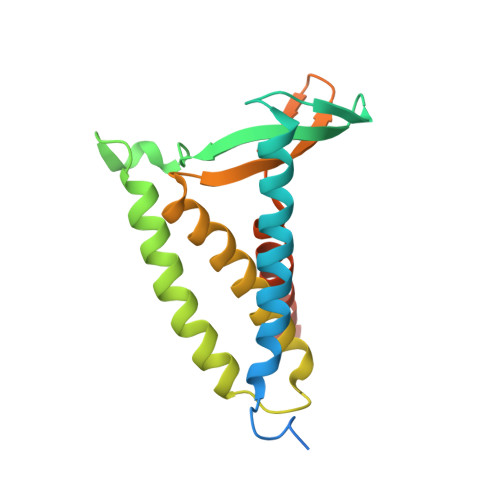
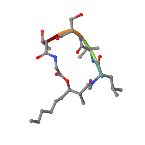
5DIR

membrane protein at 2.8 Angstroms


Experimental Data Snapshot

  • Method: X-RAY DIFFRACTION
  • Resolution: 2.80 Å
  • R-Value Free: 0.245 
  • R-Value Work: 0.219 
  • R-Value Observed: 0.221 

wwPDB Validation   3D Report Full Report


Ligand Structure Quality Assessment 


This is version 3.1 of the entry. See complete history


Literature

Structural basis of lipoprotein signal peptidase II action and inhibition by the antibiotic globomycin.

Vogeley, L.El Arnaout, T.Bailey, J.Stansfeld, P.J.Boland, C.Caffrey, M.

(2016) Science 351: 876-880

  • DOI: https://doi.org/10.1126/science.aad3747

  • PubMed Abstract: 

    With functions that range from cell envelope structure to signal transduction and transport, lipoproteins constitute 2 to 3% of bacterial genomes and play critical roles in bacterial physiology, pathogenicity, and antibiotic resistance. Lipoproteins are synthesized with a signal peptide securing them to the cytoplasmic membrane with the lipoprotein domain in the periplasm or outside the cell. Posttranslational processing requires a signal peptidase II (LspA) that removes the signal peptide. Here, we report the crystal structure of LspA from Pseudomonas aeruginosa complexed with the antimicrobial globomycin at 2.8 angstrom resolution. Mutagenesis studies identify LspA as an aspartyl peptidase. In an example of molecular mimicry, globomycin appears to inhibit by acting as a noncleavable peptide that sterically blocks the active site. This structure should inform rational antibiotic drug discovery.


  • Organizational Affiliation

    School of Medicine and School of Biochemistry and Immunology, Trinity College Dublin, Dublin, Ireland.


Macromolecules
Find similar proteins by:  (by identity cutoff)  |  3D Structure
Entity ID: 1
MoleculeChains Sequence LengthOrganismDetailsImage
Lipoprotein signal peptidase
A, B, C, D
188Pseudomonas aeruginosa PAO1Mutation(s): 0 
Gene Names: lspAlsPA4559
EC: 3.4.23.36
Membrane Entity: Yes 
UniProt
Find proteins for Q9HVM5 (Pseudomonas aeruginosa (strain ATCC 15692 / DSM 22644 / CIP 104116 / JCM 14847 / LMG 12228 / 1C / PRS 101 / PAO1))
Explore Q9HVM5 
Go to UniProtKB:  Q9HVM5
Entity Groups  
Sequence Clusters30% Identity50% Identity70% Identity90% Identity95% Identity100% Identity
UniProt GroupQ9HVM5
Sequence Annotations
Expand
  • Reference Sequence

Find similar proteins by:  Sequence   |   3D Structure  

Entity ID: 2
MoleculeChains Sequence LengthOrganismDetailsImage
Globomycin
E, F, G, H
5synthetic constructMutation(s): 0 
Sequence Annotations
Expand
  • Reference Sequence
Small Molecules
Ligands 1 Unique
IDChains Name / Formula / InChI Key2D Diagram3D Interactions
OLC
Query on OLC

Download Ideal Coordinates CCD File 
AA [auth D]
BA [auth D]
CA [auth D]
DA [auth D]
EA [auth F]
AA [auth D],
BA [auth D],
CA [auth D],
DA [auth D],
EA [auth F],
FA [auth F],
I [auth A],
J [auth A],
K [auth A],
L [auth A],
M [auth A],
N [auth A],
O [auth B],
P [auth B],
Q [auth B],
R [auth B],
S [auth B],
T [auth B],
U [auth B],
V [auth C],
W [auth C],
X [auth C],
Y [auth C],
Z [auth D]
(2R)-2,3-dihydroxypropyl (9Z)-octadec-9-enoate
C21 H40 O4
RZRNAYUHWVFMIP-GDCKJWNLSA-N
Modified Residues  3 Unique
IDChains TypeFormula2D DiagramParent
ALO
Query on ALO
E, F, G, H
L-PEPTIDE LINKINGC4 H9 N O3THR
IIL
Query on IIL
E, F, G, H
L-PEPTIDE LINKINGC6 H13 N O2ILE
MLE
Query on MLE
E, F, G, H
L-PEPTIDE LINKINGC7 H15 N O2LEU
Biologically Interesting Molecules (External Reference) 1 Unique
Experimental Data & Validation

Experimental Data

  • Method: X-RAY DIFFRACTION
  • Resolution: 2.80 Å
  • R-Value Free: 0.245 
  • R-Value Work: 0.219 
  • R-Value Observed: 0.221 
  • Space Group: C 1 2 1
Unit Cell:
Length ( Å )Angle ( ˚ )
a = 112.48α = 90
b = 105.88β = 96.92
c = 85.39γ = 90
Software Package:
Software NamePurpose
PHENIXrefinement
XDSdata reduction
XSCALEdata scaling
PHENIXphasing

Structure Validation

View Full Validation Report


Ligand Structure Quality Assessment 


Entry History & Funding Information

Deposition Data


Funding OrganizationLocationGrant Number
Science Foundation IrelandIreland12/IA/1255

Revision History  (Full details and data files)

  • Version 1.0: 2016-03-02
    Type: Initial release
  • Version 1.1: 2016-03-09
    Changes: Database references
  • Version 2.0: 2019-06-19
    Changes: Advisory, Atomic model, Data collection, Database references, Derived calculations, Polymer sequence, Source and taxonomy, Structure summary
  • Version 2.1: 2019-08-28
    Changes: Data collection, Structure summary
  • Version 3.0: 2023-11-15
    Changes: Atomic model, Data collection, Database references, Derived calculations, Refinement description
  • Version 3.1: 2024-10-09
    Changes: Structure summary