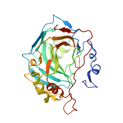
Tracking solvent and protein movement during CO2 release in carbonic anhydrase II crystals
Kim, C.U., Song, H., Avvaru, B.S., Gruner, S.M., Park, S., McKenna, R.(2016) Proc Natl Acad Sci U S A 113: 5257-5262
- PubMed: 27114542
- DOI: https://doi.org/10.1073/pnas.1520786113
- Primary Citation of Related Structures:
5DSK, 5DSL, 5DSM, 5DSO, 5DSP, 5DSQ, 5DSR, 5YUI, 5YUJ, 5YUK - PubMed Abstract:
Carbonic anhydrases are mostly zinc metalloenzymes that catalyze the reversible hydration/dehydration of CO2/HCO3 (-) Previously, the X-ray crystal structures of CO2-bound holo (zinc-bound) and apo (zinc-free) human carbonic anhydrase IIs (hCA IIs) were captured at high resolution. Here, we present sequential timeframe structures of holo- [T = 0 s (CO2-bound), 50 s, 3 min, 10 min, 25 min, and 1 h] and apo-hCA IIs [T = 0 s, 50 s, 3 min, and 10 min] during the "slow" release of CO2 Two active site waters, WDW (deep water) and WDW' (this study), replace the vacated space created on CO2 release, and another water, WI (intermediate water), is seen to translocate to the proton wire position W1. In addition, on the rim of the active site pocket, a water W2' (this study), in close proximity to residue His64 and W2, gradually exits the active site, whereas His64 concurrently rotates from pointing away ("out") to pointing toward ("in") active site rotameric conformation. This study provides for the first time, to our knowledge, structural "snapshots" of hCA II intermediate states during the formation of the His64-mediated proton wire that is induced as CO2 is released. Comparison of the holo- and apo-hCA II structures shows that the solvent network rearrangements require the presence of the zinc ion.
Organizational Affiliation:
Cornell High Energy Synchrotron Source, Cornell University, Ithaca, NY 14853; Department of Physics, Ulsan National Institute of Science and Technology, Ulsan 44919, Republic of Korea; cukim@unist.ac.kr psy@ssu.ac.kr rmckenna@ufl.edu.


