5ECP

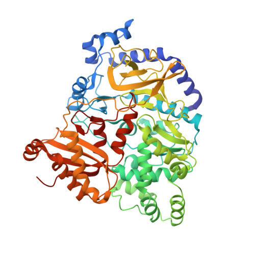
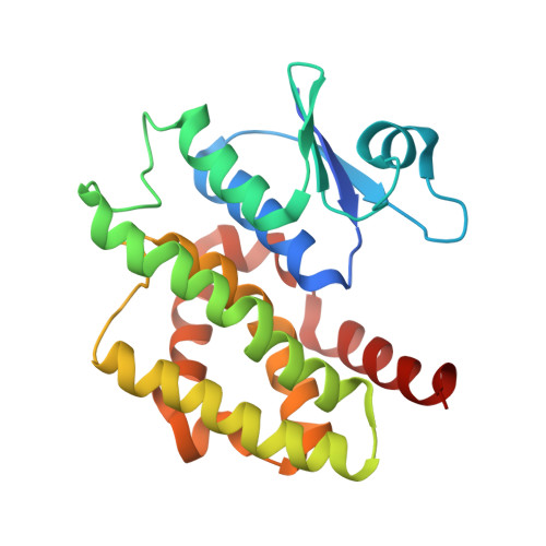
Crystal Structure of FIN219-FIP1 complex with JA, MET and ATP


Experimental Data Snapshot

  • Method: X-RAY DIFFRACTION
  • Resolution: 2.25 Å
  • R-Value Free: 0.208 
  • R-Value Work: 0.185 
  • R-Value Observed: 0.185 

Starting Model: experimental
View more details

wwPDB Validation   3D Report Full Report


Ligand Structure Quality Assessment 


This is version 1.3 of the entry. See complete history


Literature

Structural basis of jasmonate-amido synthetase FIN219 in complex with glutathione S-transferase FIP1 during the JA signal regulation

Chen, C.Y.Ho, S.S.Kuo, T.Y.Hsieh, H.L.Cheng, Y.S.

(2017) Proc Natl Acad Sci U S A 114: E1815-E1824

  • DOI: https://doi.org/10.1073/pnas.1609980114
  • Primary Citation of Related Structures:  
    5ECH, 5ECI, 5ECK, 5ECL, 5ECM, 5ECN, 5ECO, 5ECP, 5ECQ, 5ECR, 5ECS, 5GZZ

  • PubMed Abstract: 

    Far-red (FR) light-coupled jasmonate (JA) signaling is necessary for plant defense and development. FR insensitive 219 (FIN219) is a member of the Gretchen Hagen 3 (GH3) family of proteins in Arabidopsis and belongs to the adenylate-forming family of enzymes. It directly controls biosynthesis of jasmonoyl-isoleucine in JA-mediated defense responses and interacts with FIN219-interacting protein 1 (FIP1) under FR light conditions. FIN219 and FIP1 are involved in FR light signaling and are regulators of the interplay between light and JA signaling. However, how their interactions affect plant physiological functions remains unclear. Here, we demonstrate the crystal structures of FIN219-FIP1 while binding with substrates at atomic resolution. Our results show an unexpected FIN219 conformation and demonstrate various differences between this protein and other members of the GH3 family. We show that the rotated C-terminal domain of FIN219 alters ATP binding and the core structure of the active site. We further demonstrate that this unique FIN219-FIP1 structure is crucial for increasing FIN219 activity and determines the priority of substrate binding. We suggest that the increased FIN219 activity resulting from the complex form, a conformation for domain switching, allows FIN219 to switch to its high-affinity mode and thereby enhances JA signaling under continuous FR light conditions.


  • Organizational Affiliation

    Institute of Plant Biology, National Taiwan University, Taipei 10617, Taiwan.


Macromolecules
Find similar proteins by:  (by identity cutoff)  |  3D Structure
Entity ID: 1
MoleculeChains Sequence LengthOrganismDetailsImage
Jasmonic acid-amido synthetase JAR1
A, D
575Arabidopsis thalianaMutation(s): 0 
Gene Names: JAR1FIN219At2g46370F11C10.6
EC: 6.3.2 (PDB Primary Data), 6.3.2.52 (UniProt)
UniProt
Find proteins for Q9SKE2 (Arabidopsis thaliana)
Explore Q9SKE2 
Go to UniProtKB:  Q9SKE2
Entity Groups  
Sequence Clusters30% Identity50% Identity70% Identity90% Identity95% Identity100% Identity
UniProt GroupQ9SKE2
Sequence Annotations
Expand
  • Reference Sequence
Find similar proteins by:  (by identity cutoff)  |  3D Structure
Entity ID: 2
MoleculeChains Sequence LengthOrganismDetailsImage
Glutathione S-transferase U20
B, C, E, F
223Arabidopsis thalianaMutation(s): 0 
Gene Names: GSTU20FIP1At1g78370F3F9.11
EC: 2.5.1.18
UniProt
Find proteins for Q8L7C9 (Arabidopsis thaliana)
Explore Q8L7C9 
Go to UniProtKB:  Q8L7C9
Entity Groups  
Sequence Clusters30% Identity50% Identity70% Identity90% Identity95% Identity100% Identity
UniProt GroupQ8L7C9
Sequence Annotations
Expand
  • Reference Sequence
Small Molecules
Ligands 4 Unique
IDChains Name / Formula / InChI Key2D Diagram3D Interactions
ATP
Query on ATP

Download Ideal Coordinates CCD File 
I [auth A],
N [auth D]
ADENOSINE-5'-TRIPHOSPHATE
C10 H16 N5 O13 P3
ZKHQWZAMYRWXGA-KQYNXXCUSA-N
GSH
Query on GSH

Download Ideal Coordinates CCD File 
J [auth B],
K [auth C],
O [auth E],
P [auth F]
GLUTATHIONE
C10 H17 N3 O6 S
RWSXRVCMGQZWBV-WDSKDSINSA-N
JAA
Query on JAA

Download Ideal Coordinates CCD File 
G [auth A],
L [auth D]
{(1R,2R)-3-oxo-2-[(2Z)-pent-2-en-1-yl]cyclopentyl}acetic acid
C12 H18 O3
ZNJFBWYDHIGLCU-HWKXXFMVSA-N
MET
Query on MET

Download Ideal Coordinates CCD File 
H [auth A],
M [auth D]
METHIONINE
C5 H11 N O2 S
FFEARJCKVFRZRR-BYPYZUCNSA-N
Experimental Data & Validation

Experimental Data

  • Method: X-RAY DIFFRACTION
  • Resolution: 2.25 Å
  • R-Value Free: 0.208 
  • R-Value Work: 0.185 
  • R-Value Observed: 0.185 
  • Space Group: P 1
Unit Cell:
Length ( Å )Angle ( ˚ )
a = 53.708α = 89.96
b = 53.701β = 90.16
c = 192.029γ = 113.24
Software Package:
Software NamePurpose
HKL-2000data collection
DENZOdata reduction
SCALEPACKdata scaling
PHASERphasing
PHENIXmodel building
Cootmodel building
PHENIXrefinement
PDB_EXTRACTdata extraction

Structure Validation

View Full Validation Report


Ligand Structure Quality Assessment 


Entry History 

Deposition Data

Revision History  (Full details and data files)

  • Version 1.0: 2016-11-02
    Type: Initial release
  • Version 1.1: 2017-03-08
    Changes: Database references
  • Version 1.2: 2017-03-22
    Changes: Database references
  • Version 1.3: 2023-11-08
    Changes: Data collection, Database references, Refinement description