Structural and Kinetic Studies of the Human Nudix Hydrolase MTH1 Reveal the Mechanism for Its Broad Substrate Specificity
Waz, S., Nakamura, T., Hirata, K., Koga-Ogawa, Y., Chirifu, M., Arimori, T., Tamada, T., Ikemizu, S., Nakabeppu, Y., Yamagata, Y.(2017) J Biol Chem 292: 2785-2794
- PubMed: 28035004
- DOI: https://doi.org/10.1074/jbc.M116.749713
- Primary Citation of Related Structures:
5GHI, 5GHJ, 5GHM, 5GHN, 5GHO, 5GHP, 5GHQ, 5WS7, 6ILI - PubMed Abstract:
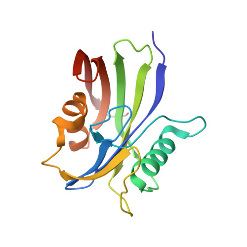
The human MutT homolog 1 (hMTH1, human NUDT1) hydrolyzes oxidatively damaged nucleoside triphosphates and is the main enzyme responsible for nucleotide sanitization. hMTH1 recently has received attention as an anticancer target because hMTH1 blockade leads to accumulation of oxidized nucleotides in the cell, resulting in mutations and death of cancer cells. Unlike Escherichia coli MutT, which shows high substrate specificity for 8-oxoguanine nucleotides, hMTH1 has broad substrate specificity for oxidized nucleotides, including 8-oxo-dGTP and 2-oxo-dATP. However, the reason for this broad substrate specificity remains unclear. Here, we determined crystal structures of hMTH1 in complex with 8-oxo-dGTP or 2-oxo-dATP at neutral pH. These structures based on high quality data showed that the base moieties of two substrates are located on the similar but not the same position in the substrate binding pocket and adopt a different hydrogen-bonding pattern, and both triphosphate moieties bind to the hMTH1 Nudix motif ( i.e. the hydrolase motif) similarly and align for the hydrolysis reaction. We also performed kinetic assays on the substrate-binding Asp-120 mutants (D120N and D120A), and determined their crystal structures in complex with the substrates. Analyses of bond lengths with high-resolution X-ray data and the relationship between the structure and enzymatic activity revealed that hMTH1 recognizes the different oxidized nucleotides via an exchange of the protonation state at two neighboring aspartate residues (Asp-119 and Asp-120) in its substrate binding pocket. To our knowledge, this mechanism of broad substrate recognition by enzymes has not been reported previously and may have relevance for anticancer drug development strategies targeting hMTH1.
Organizational Affiliation:
From the Graduate School of Pharmaceutical Sciences, Kumamoto University, Kumamoto 862-0973.


