5GZA

protein O-mannose kinase


Experimental Data Snapshot

  • Method: X-RAY DIFFRACTION
  • Resolution: 2.00 Å
  • R-Value Free: 0.218 
  • R-Value Work: 0.197 
  • R-Value Observed: 0.203 

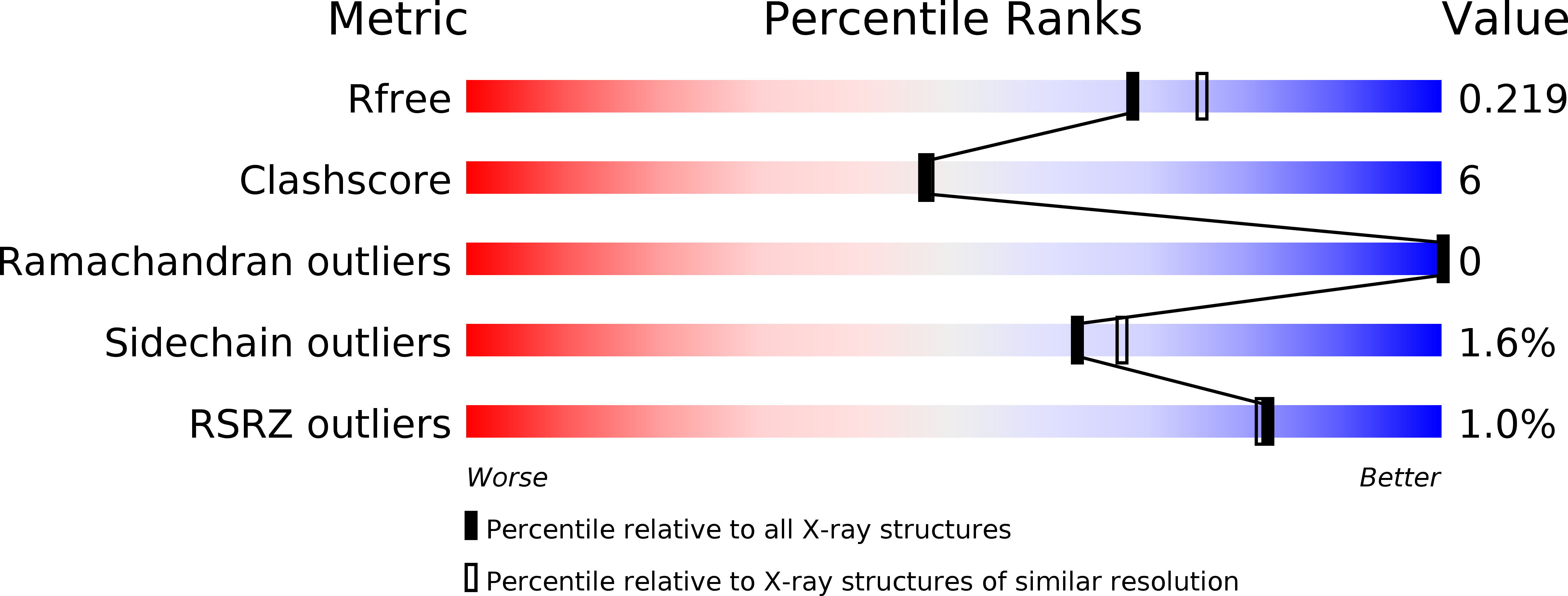
wwPDB Validation   3D Report Full Report


Ligand Structure Quality Assessment 


This is version 2.1 of the entry. See complete history


Literature

Structure of protein O-mannose kinase reveals a unique active site architecture

Zhu, Q.Venzke, D.Walimbe, A.S.Anderson, M.E.Fu, Q.Kinch, L.N.Wang, W.Chen, X.Grishin, N.V.Huang, N.Yu, L.Dixon, J.E.Campbell, K.P.Xiao, J.

(2016) Elife 5

  • DOI: https://doi.org/10.7554/eLife.22238
  • Primary Citation of Related Structures:  
    5GZA

  • PubMed Abstract: 

    The 'pseudokinase' SgK196 is a protein O-mannose kinase (POMK) that catalyzes an essential phosphorylation step during biosynthesis of the laminin-binding glycan on α-dystroglycan. However, the catalytic mechanism underlying this activity remains elusive. Here we present the crystal structure of Danio rerio POMK in complex with Mg 2+ ions, ADP, aluminum fluoride, and the GalNAc-β3-GlcNAc-β4-Man trisaccharide substrate, thereby providing a snapshot of the catalytic transition state of this unusual kinase. The active site of POMK is established by residues located in non-canonical positions and is stabilized by a disulfide bridge. GalNAc-β3-GlcNAc-β4-Man is recognized by a surface groove, and the GalNAc-β3-GlcNAc moiety mediates the majority of interactions with POMK. Expression of various POMK mutants in POMK knockout cells further validated the functional requirements of critical residues. Our results provide important insights into the ability of POMK to function specifically as a glycan kinase, and highlight the structural diversity of the human kinome.


  • Organizational Affiliation

    The State Key Laboratory of Protein and Plant Gene Research, School of Life Sciences, Peking University, Beijing, China.


Macromolecules
Find similar proteins by:  (by identity cutoff)  |  3D Structure
Entity ID: 1
MoleculeChains Sequence LengthOrganismDetailsImage
Protein O-mannose kinase290Danio rerioMutation(s): 0 
Gene Names: pomksgk196
EC: 2.7.1.183
UniProt
Find proteins for Q5U3W1 (Danio rerio)
Explore Q5U3W1 
Go to UniProtKB:  Q5U3W1
Entity Groups  
Sequence Clusters30% Identity50% Identity70% Identity90% Identity95% Identity100% Identity
UniProt GroupQ5U3W1
Sequence Annotations
Expand
  • Reference Sequence
Oligosaccharides

Help

Entity ID: 2
MoleculeChains Length2D Diagram Glycosylation3D Interactions
2-acetamido-2-deoxy-beta-D-galactopyranose-(1-3)-2-acetamido-2-deoxy-beta-D-glucopyranose-(1-4)-alpha-D-mannopyranose
B
3N/A
Glycosylation Resources
GlyTouCan:  G82715NG
GlyCosmos:  G82715NG
Small Molecules
Ligands 4 Unique
IDChains Name / Formula / InChI Key2D Diagram3D Interactions
ADP
Query on ADP

Download Ideal Coordinates CCD File 
C [auth A]ADENOSINE-5'-DIPHOSPHATE
C10 H15 N5 O10 P2
XTWYTFMLZFPYCI-KQYNXXCUSA-N
ZZ1
Query on ZZ1

Download Ideal Coordinates CCD File 
G [auth A]4-METHYL-2H-CHROMEN-2-ONE
C10 H8 O2
PSGQCCSGKGJLRL-UHFFFAOYSA-N
AF3
Query on AF3

Download Ideal Coordinates CCD File 
F [auth A]ALUMINUM FLUORIDE
Al F3
KLZUFWVZNOTSEM-UHFFFAOYSA-K
MG
Query on MG

Download Ideal Coordinates CCD File 
D [auth A],
E [auth A]
MAGNESIUM ION
Mg
JLVVSXFLKOJNIY-UHFFFAOYSA-N
Experimental Data & Validation

Experimental Data

  • Method: X-RAY DIFFRACTION
  • Resolution: 2.00 Å
  • R-Value Free: 0.218 
  • R-Value Work: 0.197 
  • R-Value Observed: 0.203 
  • Space Group: P 32
Unit Cell:
Length ( Å )Angle ( ˚ )
a = 70.55α = 90
b = 70.55β = 90
c = 66.935γ = 120
Software Package:
Software NamePurpose
SCALEPACKdata scaling
SOLVEphasing
PHENIXrefinement
PDB_EXTRACTdata extraction

Structure Validation

View Full Validation Report


Ligand Structure Quality Assessment 


Entry History 

Deposition Data

  • Released Date: 2016-12-07 
  • Deposition Author(s): Xiao, J.

Revision History  (Full details and data files)

  • Version 1.0: 2016-12-07
    Type: Initial release
  • Version 2.0: 2020-07-29
    Type: Remediation
    Reason: Carbohydrate remediation
    Changes: Advisory, Atomic model, Data collection, Derived calculations, Structure summary
  • Version 2.1: 2024-11-06
    Changes: Data collection, Database references, Derived calculations, Structure summary