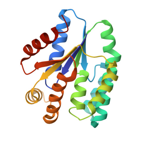
5H56

ADP and dTDP bound Crystal structure of thymidylate kinase (aq_969) from Aquifex Aeolicus VF5


Experimental Data Snapshot

  • Method: X-RAY DIFFRACTION
  • Resolution: 1.70 Å
  • R-Value Free: 0.185 
  • R-Value Work: 0.156 
  • R-Value Observed: 0.157 

Starting Model: experimental
View more details

wwPDB Validation   3D Report Full Report


Ligand Structure Quality Assessment 


This is version 1.2 of the entry. See complete history


Literature

Structural studies of a hyperthermophilic thymidylate kinase enzyme reveal conformational substates along the reaction coordinate

Biswas, A.Shukla, A.Chaudhary, S.K.Santhosh, R.Jeyakanthan, J.Sekar, K.

(2017) FEBS J 284: 2527-2544

  • DOI: https://doi.org/10.1111/febs.14140
  • Primary Citation of Related Structures:  
    4S2E, 4S35, 5H56, 5H5B, 5H5K, 5XAI, 5XB2, 5XB3, 5XB5, 5XBH

  • PubMed Abstract: 

    Thymidylate kinase (TMK) is a key enzyme which plays an important role in DNA synthesis. It belongs to the family of nucleoside monophosphate kinases, several of which undergo structure-encoded conformational changes to perform their function. However, the absence of three-dimensional structures for all the different reaction intermediates of a single TMK homolog hinders a clear understanding of its functional mechanism. We herein report the different conformational states along the reaction coordinate of a hyperthermophilic TMK from Aquifex aeolicus, determined via X-ray diffraction and further validated through normal-mode studies. The analyses implicate an arginine residue in the Lid region in catalysis, which was confirmed through site-directed mutagenesis and subsequent enzyme assays on the wild-type protein and mutants. Furthermore, the enzyme was found to exhibit broad specificity toward phosphate group acceptor nucleotides. Our comprehensive analyses of the conformational landscape of TMK, together with associated biochemical experiments, provide insights into the mechanistic details of TMK-driven catalysis, for example, the order of substrate binding and the reaction mechanism for phosphate transfer. Such a study has utility in the design of potent inhibitors for these enzymes. Structural data are available in the PDB under the accession numbers 2PBR, 4S2E, 5H5B, 5XAI, 4S35, 5XB2, 5H56, 5XB3, 5H5K, 5XB5, and 5XBH.


  • Organizational Affiliation

    Department of Physics, Indian Institute of Science, Bangalore, India.


Macromolecules
Find similar proteins by:  (by identity cutoff)  |  3D Structure
Entity ID: 1
MoleculeChains Sequence LengthOrganismDetailsImage
Thymidylate kinase
A, B
195Aquifex aeolicus VF5Mutation(s): 0 
Gene Names: tmkaq_969
EC: 2.7.4.9
UniProt
Find proteins for O67099 (Aquifex aeolicus (strain VF5))
Explore O67099 
Go to UniProtKB:  O67099
Entity Groups  
Sequence Clusters30% Identity50% Identity70% Identity90% Identity95% Identity100% Identity
UniProt GroupO67099
Sequence Annotations
Expand
  • Reference Sequence
Small Molecules
Ligands 5 Unique
IDChains Name / Formula / InChI Key2D Diagram3D Interactions
ADP
Query on ADP

Download Ideal Coordinates CCD File 
C [auth A],
K [auth B]
ADENOSINE-5'-DIPHOSPHATE
C10 H15 N5 O10 P2
XTWYTFMLZFPYCI-KQYNXXCUSA-N
TYD
Query on TYD

Download Ideal Coordinates CCD File 
D [auth A],
L [auth B]
THYMIDINE-5'-DIPHOSPHATE
C10 H16 N2 O11 P2
UJLXYODCHAELLY-XLPZGREQSA-N
MPD
Query on MPD

Download Ideal Coordinates CCD File 
J [auth A],
P [auth B]
(4S)-2-METHYL-2,4-PENTANEDIOL
C6 H14 O2
SVTBMSDMJJWYQN-YFKPBYRVSA-N
CA
Query on CA

Download Ideal Coordinates CCD File 
H [auth A],
I [auth A]
CALCIUM ION
Ca
BHPQYMZQTOCNFJ-UHFFFAOYSA-N
MG
Query on MG

Download Ideal Coordinates CCD File 
E [auth A]
F [auth A]
G [auth A]
M [auth B]
N [auth B]
E [auth A],
F [auth A],
G [auth A],
M [auth B],
N [auth B],
O [auth B]
MAGNESIUM ION
Mg
JLVVSXFLKOJNIY-UHFFFAOYSA-N
Experimental Data & Validation

Experimental Data

  • Method: X-RAY DIFFRACTION
  • Resolution: 1.70 Å
  • R-Value Free: 0.185 
  • R-Value Work: 0.156 
  • R-Value Observed: 0.157 
  • Space Group: P 1 21 1
Unit Cell:
Length ( Å )Angle ( ˚ )
a = 42.482α = 90
b = 62.414β = 91.02
c = 76.144γ = 90
Software Package:
Software NamePurpose
REFMACrefinement
HKL-2000data reduction
HKL-2000data scaling
PHASERphasing

Structure Validation

View Full Validation Report


Ligand Structure Quality Assessment 


Entry History 

Deposition Data

Revision History  (Full details and data files)

  • Version 1.0: 2017-06-28
    Type: Initial release
  • Version 1.1: 2017-08-30
    Changes: Database references
  • Version 1.2: 2023-11-08
    Changes: Data collection, Database references, Derived calculations, Refinement description