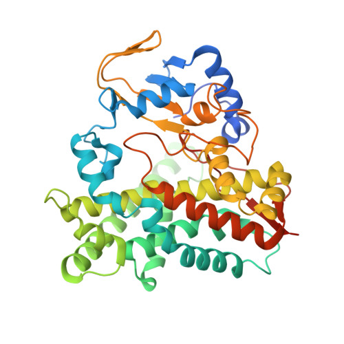
5OFQ

Crystal structure of substrate-free CYP109A2 from Bacillus megaterium


Experimental Data Snapshot

  • Method: X-RAY DIFFRACTION
  • Resolution: 2.70 Å
  • R-Value Free: 0.275 
  • R-Value Work: 0.224 
  • R-Value Observed: 0.226 

Starting Model: experimental
View more details

wwPDB Validation   3D Report Full Report


Ligand Structure Quality Assessment 


This is version 1.4 of the entry. See complete history


Literature

Biochemical and structural characterization of CYP109A2, a vitamin D3 25-hydroxylase from Bacillus megaterium.

Abdulmughni, A.Jozwik, I.K.Brill, E.Hannemann, F.Thunnissen, A.W.H.Bernhardt, R.

(2017) FEBS J 284: 3881-3894

  • DOI: https://doi.org/10.1111/febs.14276
  • Primary Citation of Related Structures:  
    5OFQ

  • PubMed Abstract: 

    Cytochrome P450 enzymes are increasingly investigated due to their potential application as biocatalysts with high regio- and/or stereo-selectivity and under mild conditions. Vitamin D 3 (VD 3 ) metabolites are of pharmaceutical importance and are applied for the treatment of VD 3 deficiency and other disorders. However, the chemical synthesis of VD 3 derivatives shows low specificity and low yields. In this study, cytochrome P450 CYP109A2 from Bacillus megaterium DSM319 was expressed, purified, and shown to oxidize VD 3 with high regio-selectivity. The in vitro conversion, using cytochrome P450 reductase (BmCPR) and ferredoxin (Fdx2) from the same strain, showed typical Michaelis-Menten reaction kinetics. A whole-cell system in B. megaterium overexpressing CYP109A2 reached 76 ± 5% conversion after 24 h and allowed to identify the main product by NMR analysis as 25-hydroxylated VD 3 . Product yield amounted to 54.9 mg·L -1 ·day -1 , rendering the established whole-cell system as a highly promising biocatalytic route for the production of this valuable metabolite. The crystal structure of substrate-free CYP109A2 was determined at 2.7 Å resolution, displaying an open conformation. Structural analysis predicts that CYP109A2 uses a highly similar set of residues for VD 3 binding as the related VD 3 hydroxylases CYP109E1 from B. megaterium and CYP107BR1 (Vdh) from Pseudonocardia autotrophica. However, the folds and sequences of the BC loops in these three P450s are highly divergent, leading to differences in the shape and apolar/polar surface distribution of their active site pockets, which may account for the observed differences in substrate specificity and the regio-selectivity of VD 3 hydroxylation. The atomic coordinates and structure factors have been deposited in the Protein Data Bank with accession code 5OFQ (substrate-free CYP109A2). Cytochrome P450 monooxygenase CYP109A2, EC 1.14.14.1, UniProt ID: D5DF88, Ferredoxin, UniProt ID: D5DFQ0, cytochrome P450 reductase, EC 1.8.1.2, UniProt ID: D5DGX1.


  • Organizational Affiliation

    Department of Biochemistry, Saarland University, Saarbrücken, Germany.


Macromolecules
Find similar proteins by:  (by identity cutoff)  |  3D Structure
Entity ID: 1
MoleculeChains Sequence LengthOrganismDetailsImage
Cytochrome P450
A, B, C, D
409Priestia megaterium DSM 319Mutation(s): 0 
Gene Names: BMD_2035
EC: 1.14
UniProt
Find proteins for D5DF88 (Priestia megaterium (strain DSM 319 / IMG 1521))
Explore D5DF88 
Go to UniProtKB:  D5DF88
Entity Groups  
Sequence Clusters30% Identity50% Identity70% Identity90% Identity95% Identity100% Identity
UniProt GroupD5DF88
Sequence Annotations
Expand
  • Reference Sequence
Small Molecules
Ligands 3 Unique
IDChains Name / Formula / InChI Key2D Diagram3D Interactions
HEM
Query on HEM

Download Ideal Coordinates CCD File 
E [auth A],
J [auth B],
O [auth C],
P [auth D]
PROTOPORPHYRIN IX CONTAINING FE
C34 H32 Fe N4 O4
KABFMIBPWCXCRK-RGGAHWMASA-L
1PE
Query on 1PE

Download Ideal Coordinates CCD File 
F [auth A],
G [auth A],
K [auth B],
M [auth C],
N [auth C]
PENTAETHYLENE GLYCOL
C10 H22 O6
JLFNLZLINWHATN-UHFFFAOYSA-N
SO4
Query on SO4

Download Ideal Coordinates CCD File 
H [auth A],
I [auth A],
L [auth B]
SULFATE ION
O4 S
QAOWNCQODCNURD-UHFFFAOYSA-L
Experimental Data & Validation

Experimental Data

  • Method: X-RAY DIFFRACTION
  • Resolution: 2.70 Å
  • R-Value Free: 0.275 
  • R-Value Work: 0.224 
  • R-Value Observed: 0.226 
  • Space Group: P 21 21 21
Unit Cell:
Length ( Å )Angle ( ˚ )
a = 77.125α = 90
b = 155.534β = 90
c = 158.151γ = 90
Software Package:
Software NamePurpose
EDNAdata collection
XDSdata reduction
Aimlessdata scaling
PHASERphasing
PHENIXrefinement
PDB_EXTRACTdata extraction

Structure Validation

View Full Validation Report


Ligand Structure Quality Assessment 


Entry History & Funding Information

Deposition Data


Funding OrganizationLocationGrant Number
European CommissionNetherlands289217

Revision History  (Full details and data files)

  • Version 1.0: 2017-09-27
    Type: Initial release
  • Version 1.1: 2017-10-04
    Changes: Database references
  • Version 1.2: 2017-11-29
    Changes: Database references
  • Version 1.3: 2018-01-24
    Changes: Source and taxonomy
  • Version 1.4: 2024-01-17
    Changes: Data collection, Database references, Refinement description