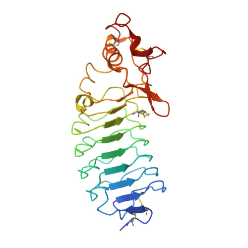
5UFB

Crystal Structure of Variable Lymphocyte Receptor (VLR) Tn4-22 (Apo)


Experimental Data Snapshot

  • Method: X-RAY DIFFRACTION
  • Resolution: 1.84 Å
  • R-Value Free: 0.213 
  • R-Value Work: 0.206 
  • R-Value Observed: 0.207 

Starting Model: experimental
View more details

wwPDB Validation   3D Report Full Report


This is version 1.4 of the entry. See complete history


Literature

Structural Insights into VLR Fine Specificity for Blood Group Carbohydrates.

Collins, B.C.Gunn, R.J.McKitrick, T.R.Cummings, R.D.Cooper, M.D.Herrin, B.R.Wilson, I.A.

(2017) Structure 25: 1667-1678.e4

  • DOI: https://doi.org/10.1016/j.str.2017.09.003
  • Primary Citation of Related Structures:  
    5UEI, 5UF1, 5UF4, 5UFB, 5UFC, 5UFD, 5UFF

  • PubMed Abstract: 

    High-quality reagents to study and detect glycans with high specificity for research and clinical applications are severely lacking. Here, we structurally and functionally characterize several variable lymphocyte receptor (VLR)-based antibodies from lampreys immunized with O erythrocytes that specifically recognize the blood group H-trisaccharide type II antigen. Glycan microarray analysis and biophysical data reveal that these VLRs exhibit greater specificity for H-trisaccharide compared with the plant lectin UEA-1, which is widely used in blood typing. Among these antibodies, O13 exhibits superior specificity for H-trisaccharide, the basis for which is revealed by comparative analysis of high-resolution VLR:glycan crystal structures. Using a structure-guided approach, we designed an O13 mutant with further enhanced specificity for H-trisaccharide. These insights into glycan recognition by VLRs suggest that lampreys can produce highly specific glycan antibodies, and are a valuable resource for the production of next-generation glycan reagents for biological and biomedical research and as diagnostics and therapeutics.


  • Organizational Affiliation

    Department of Integrative Structural and Computational Biology and the Skaggs Institute for Chemical Biology, The Scripps Research Institute, La Jolla, CA 92037, USA.


Macromolecules
Find similar proteins by:  (by identity cutoff)  |  3D Structure
Entity ID: 1
MoleculeChains Sequence LengthOrganismDetailsImage
Tn4-22248Petromyzon marinusMutation(s): 0 
Entity Groups  
Sequence Clusters30% Identity50% Identity70% Identity90% Identity95% Identity100% Identity
Sequence Annotations
Expand
  • Reference Sequence
Experimental Data & Validation

Experimental Data

  • Method: X-RAY DIFFRACTION
  • Resolution: 1.84 Å
  • R-Value Free: 0.213 
  • R-Value Work: 0.206 
  • R-Value Observed: 0.207 
  • Space Group: I 41
Unit Cell:
Length ( Å )Angle ( ˚ )
a = 83.688α = 90
b = 83.688β = 90
c = 115.073γ = 90
Software Package:
Software NamePurpose
PHENIXrefinement
PDB_EXTRACTdata extraction
XDSdata reduction
XDSdata scaling
PHASERphasing

Structure Validation

View Full Validation Report


Entry History & Funding Information

Deposition Data


Funding OrganizationLocationGrant Number
National Institutes of Health/National Institute Of Allergy and Infectious Diseases (NIH/NIAID)United States--

Revision History  (Full details and data files)

  • Version 1.0: 2017-10-18
    Type: Initial release
  • Version 1.1: 2017-11-22
    Changes: Database references
  • Version 1.2: 2019-12-11
    Changes: Author supporting evidence
  • Version 1.3: 2023-10-04
    Changes: Data collection, Database references, Refinement description
  • Version 1.4: 2024-11-06
    Changes: Structure summary