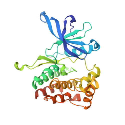
5UT3

JAK2 JH2 in complex with IKK-2 Inhibitor VI


Experimental Data Snapshot

  • Method: X-RAY DIFFRACTION
  • Resolution: 1.50 Å
  • R-Value Free: 0.170 
  • R-Value Work: 0.155 
  • R-Value Observed: 0.156 

Starting Model: experimental
View more details

wwPDB Validation   3D Report Full Report


Ligand Structure Quality Assessment 


This is version 1.4 of the entry. See complete history


Literature

Identification and Characterization of JAK2 Pseudokinase Domain Small Molecule Binders.

Puleo, D.E.Kucera, K.Hammaren, H.M.Ungureanu, D.Newton, A.S.Silvennoinen, O.Jorgensen, W.L.Schlessinger, J.

(2017) ACS Med Chem Lett 8: 618-621

  • DOI: https://doi.org/10.1021/acsmedchemlett.7b00153
  • Primary Citation of Related Structures:  
    5USY, 5USZ, 5UT0, 5UT1, 5UT2, 5UT3

  • PubMed Abstract: 

    Janus kinases (JAKs) regulate hematopoiesis via the cytokine-mediated JAK-STAT signaling pathway. JAKs contain tandem C-terminal pseudokinase (JH2) and tyrosine kinase (JH1) domains. The JAK2 pseudokinase domain adopts a protein kinase fold and, despite its pseudokinase designation, binds ATP with micromolar affinity. Recent evidence shows that displacing ATP from the JAK2 JH2 domain alters the hyperactivation state of the oncogenic JAK2 V617F protein while sparing the wild type JAK2 protein. In this study, small molecule binders of JAK2 JH2 were identified via an in vitro screen. Top hits were characterized using biophysical and structural approaches. Development of pseudokinase-selective compounds may offer novel pharmacological opportunities for treating cancers driven by JAK2 V617F and other oncogenic JAK mutants.


  • Organizational Affiliation

    Department of Pharmacology and Department of Chemistry, Yale University, New Haven, Connecticut 06520, United States.


Macromolecules
Find similar proteins by:  (by identity cutoff)  |  3D Structure
Entity ID: 1
MoleculeChains Sequence LengthOrganismDetailsImage
Tyrosine-protein kinase JAK2289Homo sapiensMutation(s): 3 
Gene Names: JAK2
EC: 2.7.10.2
UniProt & NIH Common Fund Data Resources
Find proteins for O60674 (Homo sapiens)
Explore O60674 
Go to UniProtKB:  O60674
PHAROS:  O60674
GTEx:  ENSG00000096968 
Entity Groups  
Sequence Clusters30% Identity50% Identity70% Identity90% Identity95% Identity100% Identity
UniProt GroupO60674
Sequence Annotations
Expand
  • Reference Sequence
Experimental Data & Validation

Experimental Data

  • Method: X-RAY DIFFRACTION
  • Resolution: 1.50 Å
  • R-Value Free: 0.170 
  • R-Value Work: 0.155 
  • R-Value Observed: 0.156 
  • Space Group: P 1 21 1
Unit Cell:
Length ( Å )Angle ( ˚ )
a = 44.374α = 90
b = 57.641β = 109.94
c = 60.496γ = 90
Software Package:
Software NamePurpose
PHENIXrefinement
HKL-2000data reduction
SCALEPACKdata scaling
PHASERphasing

Structure Validation

View Full Validation Report


Ligand Structure Quality Assessment 


Entry History & Funding Information

Deposition Data


Funding OrganizationLocationGrant Number
National Institutes of Health/National Institute of General Medical Sciences (NIH/NIGMS)United StatesGM007324
Gilead SciencesUnited StatesYG-003-13

Revision History  (Full details and data files)

  • Version 1.0: 2017-06-07
    Type: Initial release
  • Version 1.1: 2017-07-05
    Changes: Database references
  • Version 1.2: 2017-09-13
    Changes: Author supporting evidence
  • Version 1.3: 2020-01-01
    Changes: Author supporting evidence
  • Version 1.4: 2023-10-04
    Changes: Data collection, Database references, Refinement description