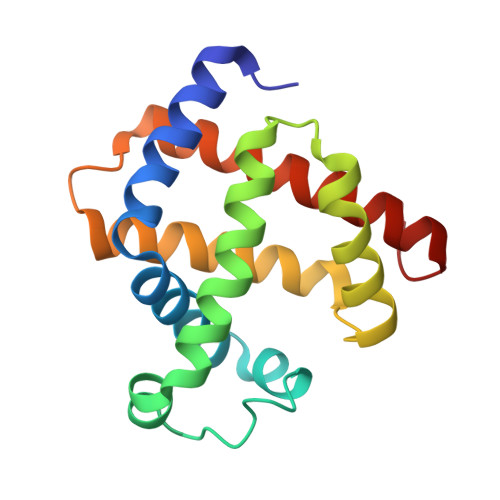
Nitrosyl Myoglobins and Their Nitrite Precursors: Crystal Structural and Quantum Mechanics and Molecular Mechanics Theoretical Investigations of Preferred Fe -NO Ligand Orientations in Myoglobin Distal Pockets.
Wang, B., Shi, Y., Tejero, J., Powell, S.M., Thomas, L.M., Gladwin, M.T., Shiva, S., Zhang, Y., Richter-Addo, G.B.(2018) Biochemistry 57: 4788-4802
- PubMed: 29999305
- DOI: https://doi.org/10.1021/acs.biochem.8b00542
- Primary Citation of Related Structures:
5UT7, 5UT8, 5UT9, 5UTA, 5UTB, 5UTC, 5UTD, 5VZN, 5VZO, 5VZP, 5VZQ, 6CF0 - PubMed Abstract:
The globular dioxygen binding heme protein myoglobin (Mb) is present in several species. Its interactions with the simple nitrogen oxides, namely, nitric oxide (NO) and nitrite, have been known for decades, but the physiological relevance has only recently become more fully appreciated. We previously reported the O-nitrito mode of binding of nitrite to ferric horse heart wild-type (wt) Mb III and human hemoglobin. We have expanded on this work and report the interactions of nitrite with wt sperm whale (sw) Mb III and its H64A, H64Q, and V68A/I107Y mutants whose dissociation constants increase in the following order: H64Q < wt < V68A/I107Y < H64A. We also report their X-ray crystal structures that reveal the O-nitrito mode of binding of nitrite to these derivatives. The Mb II -mediated reductions of nitrite to NO and structural data for the wt and mutant Mb II -NOs are described. We show that their FeNO orientations vary with distal pocket identity, with the FeNO moieties pointing toward the hydrophobic interiors when the His64 residue is present but toward the hydrophilic exterior when this His64 residue is absent in this set of mutants. This correlates with the nature of H-bonding to the bound NO ligand (nitrosyl O vs N atom). Quantum mechanics and hybrid quantum mechanics and molecular mechanics calculations help elucidate the origin of the experimentally preferred NO orientations. In a few cases, the calculations reproduce the experimentally observed orientations only when the whole protein is taken into consideration.
Organizational Affiliation:
Price Family Foundation Institute of Structural Biology and Department of Chemistry and Biochemistry , University of Oklahoma , 101 Stephenson Parkway , Norman , Oklahoma 73019 , United States.


