5XGK

Crystal structure of Arabidopsis thaliana 4-hydroxyphenylpyruvate dioxygenase (AtHPPD) complexed with its substrate 4-hydroxyphenylpyruvate acid (HPPA)


Experimental Data Snapshot

  • Method: X-RAY DIFFRACTION
  • Resolution: 2.80 Å
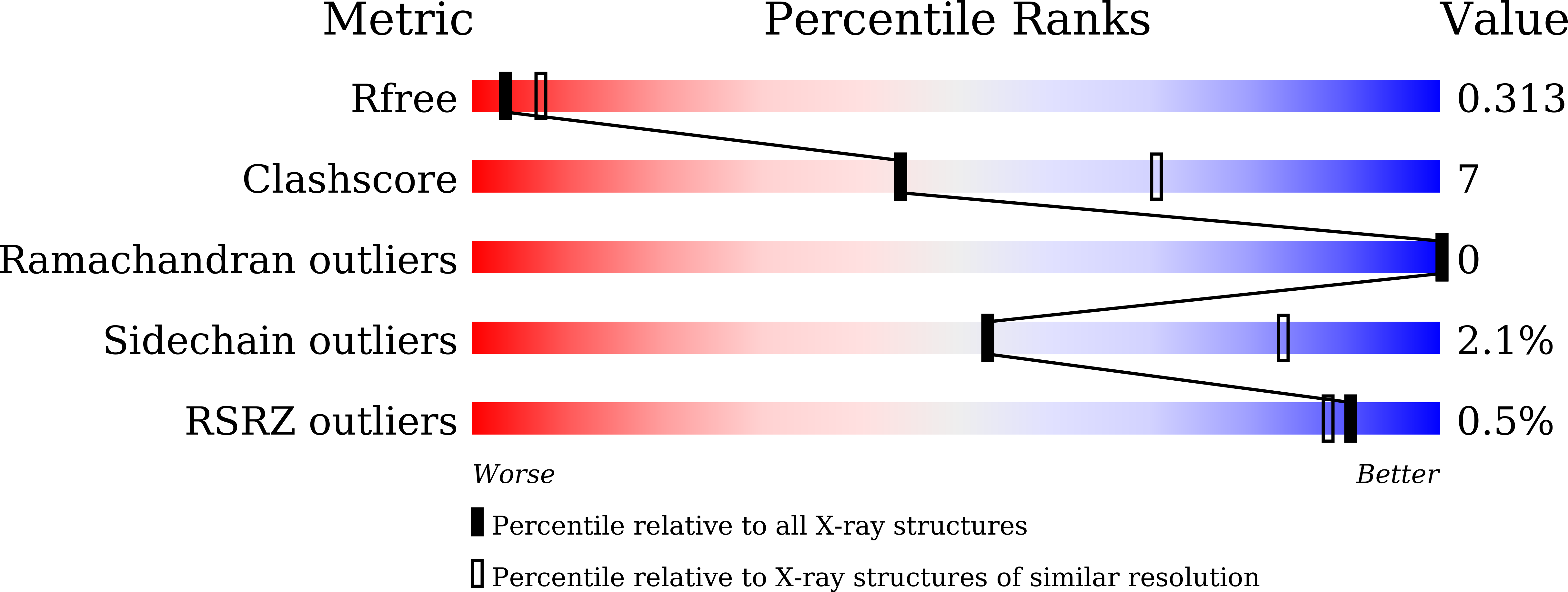
  • R-Value Free: 0.312 
  • R-Value Work: 0.258 
  • R-Value Observed: 0.261 

Starting Model: experimental
View more details

wwPDB Validation   3D Report Full Report


Ligand Structure Quality Assessment 


This is version 2.1 of the entry. See complete history


Literature

Crystal Structure of 4-Hydroxyphenylpyruvate Dioxygenase in Complex with Substrate Reveals a New Starting Point for Herbicide Discovery.

Lin, H.Y.Chen, X.Chen, J.N.Wang, D.W.Wu, F.X.Lin, S.Y.Zhan, C.G.Wu, J.W.Yang, W.C.Yang, G.F.

(2019) Research (Wash D C) 2019: 2602414-2602414

  • DOI: https://doi.org/10.34133/2019/2602414
  • Primary Citation of Related Structures:  
    5XGK, 5YY6

  • PubMed Abstract: 

    4-Hydroxyphenylpyruvate dioxygenase (HPPD) is a promising target for drug and pesticide discovery. The unknown binding mode of substrate is still a big challenge for the understanding of enzymatic reaction mechanism and novel HPPD inhibitor design. Herein, we determined the first crystal structure of Arabidopsis thaliana HPPD ( At HPPD) in complex with its natural substrate (HPPA) at a resolution of 2.80 Å. Then, combination of hybrid quantum mechanics/molecular mechanics (QM/MM) calculations confirmed that HPPA takes keto rather than enol form inside the HPPD active pocket. Subsequent site-directed mutagenesis and kinetic analysis further showed that residues (Phe424, Asn423, Glu394, Gln307, Asn282, and Ser267) played important roles in substrate binding and catalytic cycle. Structural comparison between HPPA- At HPPD and holo- At HPPD revealed that Gln293 underwent a remarkable rotation upon the HPPA binding and formed H-bond network of Ser267-Asn282-Gln307-Gln293, resulting in the transformation of HPPD from an inactive state to active state. Finally, taking the conformation change of Gln293 as a target, we proposed a new strategy of blocking the transformation of HPPD from inactive state to active state to design a novel inhibitor with K i value of 24.10 nM towards At HPPD. The inhibitor has entered into industry development as the first selective herbicide used for the weed control in sorghum field. The crystal structure of At HPPD in complex with the inhibitor (2.40 Å) confirmed the rationality of the design strategy. We believe that the present work provides a new starting point for the understanding of enzymatic reaction mechanism and the design of next generation HPPD inhibitors.


  • Organizational Affiliation

    Key Laboratory of Pesticide & Chemical Biology of Ministry of Education, International Joint Research Center for Intelligent Biosensor Technology and Health, College of Chemistry, Chemical Biology Center, Central China Normal University, Wuhan 430079, China.


Macromolecules
Find similar proteins by:  (by identity cutoff)  |  3D Structure
Entity ID: 1
MoleculeChains Sequence LengthOrganismDetailsImage
4-hydroxyphenylpyruvate dioxygenase
A, B, C, D
445Arabidopsis thalianaMutation(s): 0 
Gene Names: HPDPDS1At1g06570F12K11.9
EC: 1.13.11.27
UniProt
Find proteins for P93836 (Arabidopsis thaliana)
Explore P93836 
Go to UniProtKB:  P93836
Entity Groups  
Sequence Clusters30% Identity50% Identity70% Identity90% Identity95% Identity100% Identity
UniProt GroupP93836
Sequence Annotations
Expand
  • Reference Sequence
Small Molecules
Ligands 3 Unique
IDChains Name / Formula / InChI Key2D Diagram3D Interactions
TYF
Query on TYF

Download Ideal Coordinates CCD File 
E [auth A](2S)-2-hydroxy-3-(4-hydroxyphenyl)propanoic acid
C9 H10 O4
JVGVDSSUAVXRDY-QMMMGPOBSA-N
ACT
Query on ACT

Download Ideal Coordinates CCD File 
G [auth A],
H [auth A],
J [auth B],
M [auth D]
ACETATE ION
C2 H3 O2
QTBSBXVTEAMEQO-UHFFFAOYSA-M
FE
Query on FE

Download Ideal Coordinates CCD File 
F [auth A],
I [auth B],
K [auth C],
L [auth D]
FE (III) ION
Fe
VTLYFUHAOXGGBS-UHFFFAOYSA-N
Experimental Data & Validation

Experimental Data

  • Method: X-RAY DIFFRACTION
  • Resolution: 2.80 Å
  • R-Value Free: 0.312 
  • R-Value Work: 0.258 
  • R-Value Observed: 0.261 
  • Space Group: P 1 21 1
Unit Cell:
Length ( Å )Angle ( ˚ )
a = 95.606α = 90
b = 95.294β = 92.1
c = 98γ = 90
Software Package:
Software NamePurpose
PHENIXrefinement
HKL-2000data processing
HKL-2000data scaling
PHASERphasing
HKL-2000data reduction

Structure Validation

View Full Validation Report


Ligand Structure Quality Assessment 


Entry History & Funding Information

Deposition Data


Funding OrganizationLocationGrant Number
National Natural Science Foundation of ChinaChina21332004
National Natural Science Foundation of ChinaChina21672079

Revision History  (Full details and data files)

  • Version 1.0: 2018-09-19
    Type: Initial release
  • Version 1.1: 2019-10-02
    Changes: Data collection, Database references
  • Version 1.2: 2019-11-20
    Changes: Database references
  • Version 2.0: 2023-11-15
    Changes: Atomic model, Data collection, Database references, Derived calculations
  • Version 2.1: 2023-11-22
    Changes: Refinement description