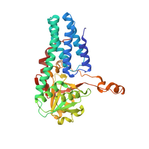
5YFS

Crystal structure of ribose-1,5-bisphosphate isomerase mutant C135S from Pyrococcus horikoshii OT3 in complex with ribose-1,5-bisphosphate


Experimental Data Snapshot

  • Method: X-RAY DIFFRACTION
  • Resolution: 2.30 Å
  • R-Value Free: 0.198 
  • R-Value Work: 0.144 
  • R-Value Observed: 0.146 

Starting Model: experimental
View more details

wwPDB Validation   3D Report Full Report


Ligand Structure Quality Assessment 


This is version 1.2 of the entry. See complete history


Literature

A presumed homologue of the regulatory subunits of eIF2B functions as ribose-1,5-bisphosphate isomerase in Pyrococcus horikoshii OT3.

Gogoi, P.Kanaujia, S.P.

(2018) Sci Rep 8: 1891-1891

  • DOI: https://doi.org/10.1038/s41598-018-20418-w
  • Primary Citation of Related Structures:  
    5YFJ, 5YFS, 5YFT, 5YFU, 5YFV, 5YFW, 5YFX, 5YG5, 5YG6, 5YG7, 5YG8, 5YG9, 5YGA

  • PubMed Abstract: 

    The homologues of the regulatory subunits of eukaryotic translation initiation factor 2B (eIF2B) are assumed to be present in archaea. Likewise, an ORF, PH0208 in Pyrococcus horikoshii OT3 have been proposed to encode one of the homologues of regulatory subunits of eIF2B. However, PH0208 protein also shares sequence similarity with a functionally non-related enzyme, ribose-1,5-bisphosphate isomerase (R15Pi), involved in conversion of ribose-1,5-bisphosphate (R15P) to ribulose-1,5-bisphosphate (RuBP) in an AMP-dependent manner. Herein, we have determined the crystal structure of PH0208 protein in order to decipher its true function. Although structurally similar to the regulatory subunits of eIF2B, the ability to bind R15P and RuBP suggests that PH0208 would function as R15Pi. Additionally, this study for the first time reports the binding sites of AMP and GMP in R15Pi. The AMP binding site in PH0208 protein clarified the role of AMP in providing structural stability to R15Pi. The binding of GMP to the 'AMP binding site' in addition to its own binding site indicates that GMP might also execute a similar function, though with less specificity. Furthermore, we have utilized the resemblance between PH0208 and the regulatory subunits of eIF2B to propose a model for the regulatory mechanism of eIF2B in eukaryotes.


  • Organizational Affiliation

    Department of Biosciences and Bioengineering, Indian Institute of Technology Guwahati, Guwahati, 781039, Assam, India.


Macromolecules
Find similar proteins by:  (by identity cutoff)  |  3D Structure
Entity ID: 1
MoleculeChains Sequence LengthOrganismDetailsImage
Ribose 1,5-bisphosphate isomerase
A, B, C
324Pyrococcus horikoshii OT3Mutation(s): 1 
Gene Names: PH0208
EC: 5.3.1.29
UniProt
Find proteins for O57947 (Pyrococcus horikoshii (strain ATCC 700860 / DSM 12428 / JCM 9974 / NBRC 100139 / OT-3))
Explore O57947 
Go to UniProtKB:  O57947
Entity Groups  
Sequence Clusters30% Identity50% Identity70% Identity90% Identity95% Identity100% Identity
UniProt GroupO57947
Sequence Annotations
Expand
  • Reference Sequence
Small Molecules
Ligands 4 Unique
IDChains Name / Formula / InChI Key2D Diagram3D Interactions
RI2 (Subject of Investigation/LOI)
Query on RI2

Download Ideal Coordinates CCD File 
D [auth A],
H [auth B],
K [auth C]
1,5-di-O-phosphono-alpha-D-ribofuranose
C5 H12 O11 P2
AAAFZMYJJHWUPN-TXICZTDVSA-N
MPD
Query on MPD

Download Ideal Coordinates CCD File 
G [auth A](4S)-2-METHYL-2,4-PENTANEDIOL
C6 H14 O2
SVTBMSDMJJWYQN-YFKPBYRVSA-N
K
Query on K

Download Ideal Coordinates CCD File 
E [auth A],
I [auth B],
L [auth C]
POTASSIUM ION
K
NPYPAHLBTDXSSS-UHFFFAOYSA-N
CL
Query on CL

Download Ideal Coordinates CCD File 
F [auth A],
J [auth B],
M [auth C]
CHLORIDE ION
Cl
VEXZGXHMUGYJMC-UHFFFAOYSA-M
Experimental Data & Validation

Experimental Data

  • Method: X-RAY DIFFRACTION
  • Resolution: 2.30 Å
  • R-Value Free: 0.198 
  • R-Value Work: 0.144 
  • R-Value Observed: 0.146 
  • Space Group: P 31 1 2
Unit Cell:
Length ( Å )Angle ( ˚ )
a = 98.65α = 90
b = 98.65β = 90
c = 256.731γ = 120
Software Package:
Software NamePurpose
HKL-3000data collection
MOSFLMdata processing
Aimlessdata scaling
PHASERphasing
REFMACrefinement
Cootmodel building
PDB_EXTRACTdata extraction

Structure Validation

View Full Validation Report


Ligand Structure Quality Assessment 


Entry History & Funding Information

Deposition Data


Funding OrganizationLocationGrant Number
Department of Biotechnology (DBT)IndiaBT/302/NE/TBP/2012 dated 07.01.2013

Revision History  (Full details and data files)

  • Version 1.0: 2018-02-14
    Type: Initial release
  • Version 1.1: 2020-07-29
    Type: Remediation
    Reason: Carbohydrate remediation
    Changes: Data collection, Derived calculations
  • Version 1.2: 2023-11-22
    Changes: Data collection, Database references, Refinement description, Structure summary