5YJ9

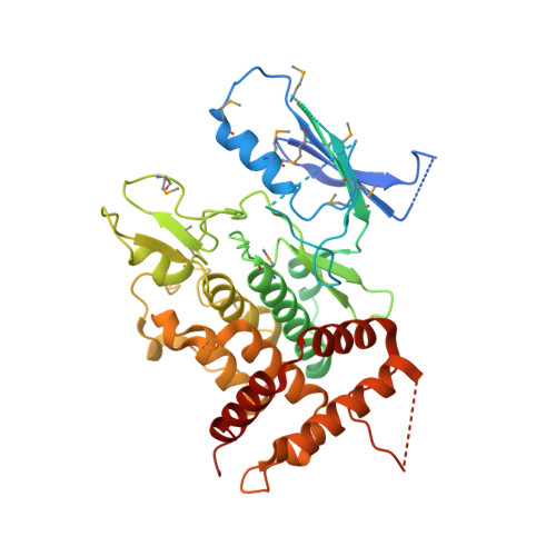
Crystal structure of Tribolium castaneum PINK1 kinase domain in complex with AMP-PNP


Experimental Data Snapshot

  • Method: X-RAY DIFFRACTION
  • Resolution: 2.53 Å
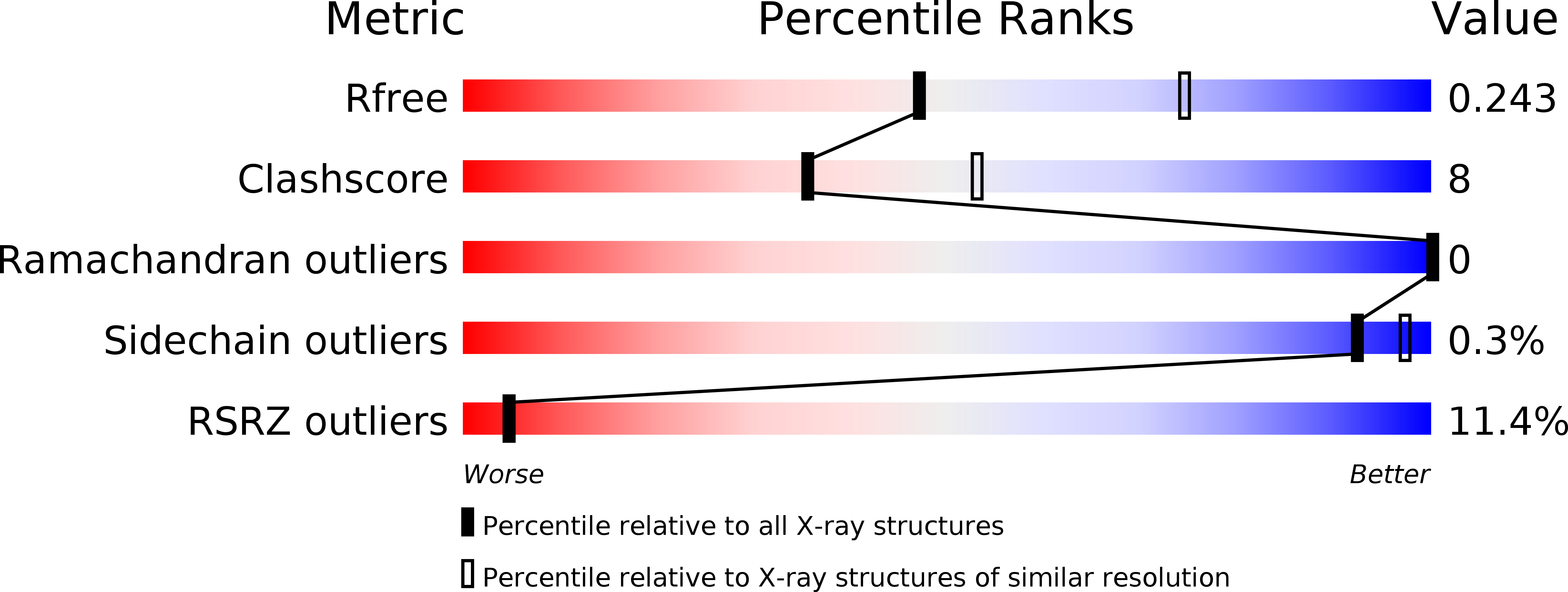
  • R-Value Free: 0.243 
  • R-Value Work: 0.229 
  • R-Value Observed: 0.230 

wwPDB Validation   3D Report Full Report


Ligand Structure Quality Assessment 


This is version 1.1 of the entry. See complete history


Literature

Structural insights into ubiquitin phosphorylation by PINK1.

Okatsu, K.Sato, Y.Yamano, K.Matsuda, N.Negishi, L.Takahashi, A.Yamagata, A.Goto-Ito, S.Mishima, M.Ito, Y.Oka, T.Tanaka, K.Fukai, S.

(2018) Sci Rep 8: 10382-10382

  • DOI: https://doi.org/10.1038/s41598-018-28656-8
  • Primary Citation of Related Structures:  
    5YJ9

  • PubMed Abstract: 

    Mutations of PTEN-induced putative kinase 1 (PINK1) and the E3 ubiquitin (Ub) ligase parkin can cause familial parkinsonism. These two proteins are essential for ubiquitylation of damaged mitochondria and subsequent degradation. PINK1 phosphorylates Ser65 of Ub and the Ub-like (UBL) domain of parkin to allosterically relieve the autoinhibition of parkin. To understand the structural mechanism of the Ub/UBL-specific phosphorylation by PINK1, we determined the crystal structure of Tribolium castaneum PINK1 kinase domain (TcPINK1) in complex with a nonhydrolyzable ATP analogue at 2.5 Å resolution. TcPINK1 consists of the N- and C-terminal lobes with the PINK1-specific extension. The ATP analogue is bound in the cleft between the N- and C-terminal lobes. The adenine ring of the ATP analogue is bound to a hydrophobic pocket, whereas the triphosphate group of the ATP analogue and two coordinated Mg ions interact with the catalytic hydrophilic residues. Comparison with protein kinases A and C (PKA and PKC, respectively) unveils a putative Ub/UBL-binding groove, which is wider than the peptide-binding groove of PKA or PKC to accommodate the globular head of Ub or UBL. Further crosslinking analyses suggested a PINK1-interacting surface of Ub. Structure-guided mutational analyses support the findings from the present structural analysis of PINK1.


  • Organizational Affiliation

    Institute for Quantitative Biosciences, The University of Tokyo, Tokyo, 113-0032, Japan.


Macromolecules
Find similar proteins by:  (by identity cutoff)  |  3D Structure
Entity ID: 1
MoleculeChains Sequence LengthOrganismDetailsImage
Serine/threonine-protein kinase PINK1, mitochondrial-like ProteinA [auth D]423Tribolium castaneumMutation(s): 4 
Gene Names: TcasGA2_TC013202
EC: 2.7.11.1
UniProt
Find proteins for D6WMX4 (Tribolium castaneum)
Explore D6WMX4 
Go to UniProtKB:  D6WMX4
Entity Groups  
Sequence Clusters30% Identity50% Identity70% Identity90% Identity95% Identity100% Identity
UniProt GroupD6WMX4
Sequence Annotations
Expand
  • Reference Sequence
Small Molecules
Modified Residues  1 Unique
IDChains TypeFormula2D DiagramParent
MSE
Query on MSE
A [auth D]L-PEPTIDE LINKINGC5 H11 N O2 SeMET
Experimental Data & Validation

Experimental Data

  • Method: X-RAY DIFFRACTION
  • Resolution: 2.53 Å
  • R-Value Free: 0.243 
  • R-Value Work: 0.229 
  • R-Value Observed: 0.230 
  • Space Group: C 1 2 1
Unit Cell:
Length ( Å )Angle ( ˚ )
a = 138.028α = 90
b = 59.79β = 101.82
c = 50.822γ = 90
Software Package:
Software NamePurpose
PHENIXrefinement
HKL-2000data reduction
HKL-2000data scaling
PHENIXphasing

Structure Validation

View Full Validation Report


Ligand Structure Quality Assessment 


Entry History & Funding Information

Deposition Data


Funding OrganizationLocationGrant Number
JSPSJapanJP15H01175
JSPSJapan24687012
JSPSJapanJP16H04750
JSPSJapanJP15J10559
CRESTJapanJPMJCR12M5

Revision History  (Full details and data files)

  • Version 1.0: 2018-07-25
    Type: Initial release
  • Version 1.1: 2024-10-23
    Changes: Data collection, Database references, Derived calculations, Structure summary