Structural Control of Nonnative Ligand Binding in Engineered Mutants of Phosphoenolpyruvate Carboxykinase.
Tang, H.Y.H., Shin, D.S., Hura, G.L., Yang, Y., Hu, X., Lightstone, F.C., McGee, M.D., Padgett, H.S., Yannone, S.M., Tainer, J.A.(2018) Biochemistry 57: 6688-6700
- PubMed: 30376300
- DOI: https://doi.org/10.1021/acs.biochem.8b00963
- Primary Citation of Related Structures:
6ASI, 6ASM, 6ASN, 6AT2, 6AT3, 6AT4 - PubMed Abstract:
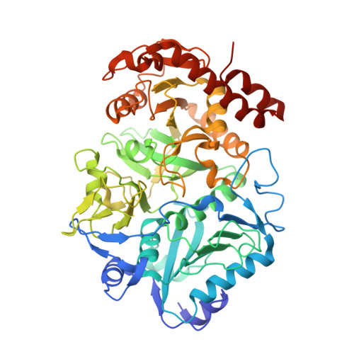
Protein engineering to alter recognition underlying ligand binding and activity has enormous potential. Here, ligand binding for Escherichia coli phosphoenolpyruvate carboxykinase (PEPCK), which converts oxaloacetate into CO 2 and phosphoenolpyruvate as the first committed step in gluconeogenesis, was engineered to accommodate alternative ligands as an exemplary system with structural information. From our identification of bicarbonate binding in the PEPCK active site at the supposed CO 2 binding site, we probed binding of nonnative ligands with three oxygen atoms arranged to resemble the bicarbonate geometry. Crystal structures of PEPCK and point mutants with bound nonnative ligands thiosulfate and methanesulfonate along with strained ATP and reoriented oxaloacetate intermediates and unexpected bicarbonate were determined and analyzed. The mutations successfully altered the bound ligand position and orientation and its specificity: mutated PEPCKs bound either thiosulfate or methanesulfonate but never both. Computational calculations predicted a methanesulfonate binding mutant and revealed that release of the active site ordered solvent exerts a strong influence on ligand binding. Besides nonnative ligand binding, one mutant altered the Mn 2+ coordination sphere: instead of the canonical octahedral ligand arrangement, the mutant in question had an only five-coordinate arrangement. From this work, critical features of ligand binding, position, and metal ion cofactor geometry required for all downstream events can be engineered with small numbers of mutations to provide insights into fundamental underpinnings of protein-ligand recognition. Through structural and computational knowledge, the combination of designed and random mutations aids in the robust design of predetermined changes to ligand binding and activity to engineer protein function.
Organizational Affiliation:
Molecular Biophysics and Integrated Bioimaging Division , Lawrence Berkeley National Laboratory , Berkeley , California 94720 , United States.


