6CXU

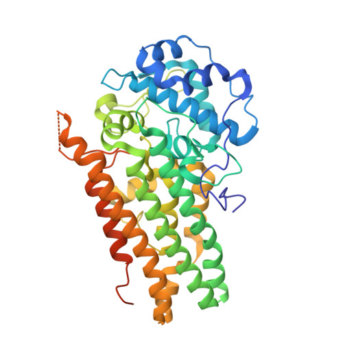
Structure of the S167H mutant of human indoleamine 2,3 dioxygenase in complex with tryptophan and cyanide


Experimental Data Snapshot

  • Method: X-RAY DIFFRACTION
  • Resolution: 2.49 Å
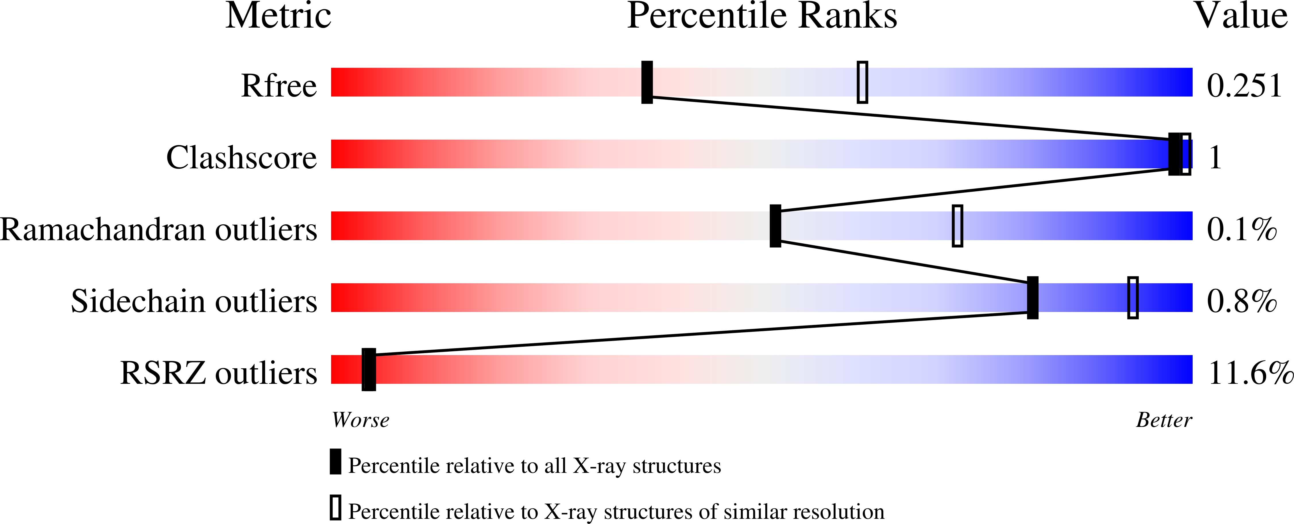
  • R-Value Free: 0.250 
  • R-Value Work: 0.201 
  • R-Value Observed: 0.204 

Starting Model: experimental
View more details

wwPDB Validation   3D Report Full Report


Ligand Structure Quality Assessment 


This is version 1.3 of the entry. See complete history


Literature

Inhibition Mechanisms of Human Indoleamine 2,3 Dioxygenase 1.

Lewis-Ballester, A.Karkashon, S.Batabyal, D.Poulos, T.L.Yeh, S.R.

(2018) J Am Chem Soc 140: 8518-8525

  • DOI: https://doi.org/10.1021/jacs.8b03691
  • Primary Citation of Related Structures:  
    6CXU, 6CXV

  • PubMed Abstract: 

    Human indoleamine 2,3-dioxygenase 1 (hIDO1) and tryptophan dioxygenase (hTDO) catalyze the same dioxygenation reaction of Trp to generate N-formyl kynurenine (NFK). They share high structural similarity, especially in the active site. However, hIDO1 possesses a unique inhibitory substrate binding site (Si) that is absent in hTDO. In addition, in hIDO1, the indoleamine group of the substrate Trp is H-bonded to S167 through a bridging water, while that in hTDO is directly H-bonded to H76. Here we show that Trp binding to the Si site or the mutation of S167 to histidine in hIDO1 retards its turnover activity and that the inhibited activity can be rescued by an effector, 3-indole ethanol (IDE). Kinetic studies reveal that the inhibited activity introduced by Trp binding to the Si site is a result of retarded recombination of the ferryl moiety with Trp epoxide to form NFK and that IDE reverses the effect by preventing Trp from binding to the Si site. In contrast, the abolished activity induced by the S167H mutation is primarily a result of ∼5000-fold reduction in the O 2 binding rate constant, possibly due to the blockage of a ligand delivery tunnel, and that IDE binding to the Si site reverses the effect by reopening the tunnel. The data offer new insights into structure-based design of hIDO1-selective inhibitors.


  • Organizational Affiliation

    Department of Physiology and Biophysics , Albert Einstein College of Medicine , Bronx , New York 10461 , United States.


Macromolecules
Find similar proteins by:  (by identity cutoff)  |  3D Structure
Entity ID: 1
MoleculeChains Sequence LengthOrganismDetailsImage
Indoleamine 2,3-dioxygenase 1
A, B
425Homo sapiensMutation(s): 1 
Gene Names: IDO1IDOINDO
EC: 1.13.11.52
UniProt & NIH Common Fund Data Resources
Find proteins for P14902 (Homo sapiens)
Explore P14902 
Go to UniProtKB:  P14902
PHAROS:  P14902
GTEx:  ENSG00000131203 
Entity Groups  
Sequence Clusters30% Identity50% Identity70% Identity90% Identity95% Identity100% Identity
UniProt GroupP14902
Sequence Annotations
Expand
  • Reference Sequence
Experimental Data & Validation

Experimental Data

  • Method: X-RAY DIFFRACTION
  • Resolution: 2.49 Å
  • R-Value Free: 0.250 
  • R-Value Work: 0.201 
  • R-Value Observed: 0.204 
  • Space Group: P 21 21 21
Unit Cell:
Length ( Å )Angle ( ˚ )
a = 86.91α = 90
b = 97.95β = 90
c = 129.34γ = 90
Software Package:
Software NamePurpose
REFMACrefinement
XDSdata reduction
Aimlessdata scaling
PHASERphasing

Structure Validation

View Full Validation Report


Ligand Structure Quality Assessment 


Entry History & Funding Information

Deposition Data


Funding OrganizationLocationGrant Number
National Institutes of Health/National Institute of General Medical Sciences (NIH/NIGMS)United StatesGM115773
National Science Foundation (NSF, United States)United StatesCHE-1404929
National Institutes of Health/National Institute of General Medical Sciences (NIH/NIGMS)United StatesGM57353

Revision History  (Full details and data files)

  • Version 1.0: 2018-06-27
    Type: Initial release
  • Version 1.1: 2018-07-25
    Changes: Data collection, Database references
  • Version 1.2: 2019-11-27
    Changes: Author supporting evidence
  • Version 1.3: 2023-10-04
    Changes: Data collection, Database references, Refinement description