6DC3

RSV prefusion F bound to RSD5 Fab


Experimental Data Snapshot

  • Method: X-RAY DIFFRACTION
  • Resolution: 3.50 Å
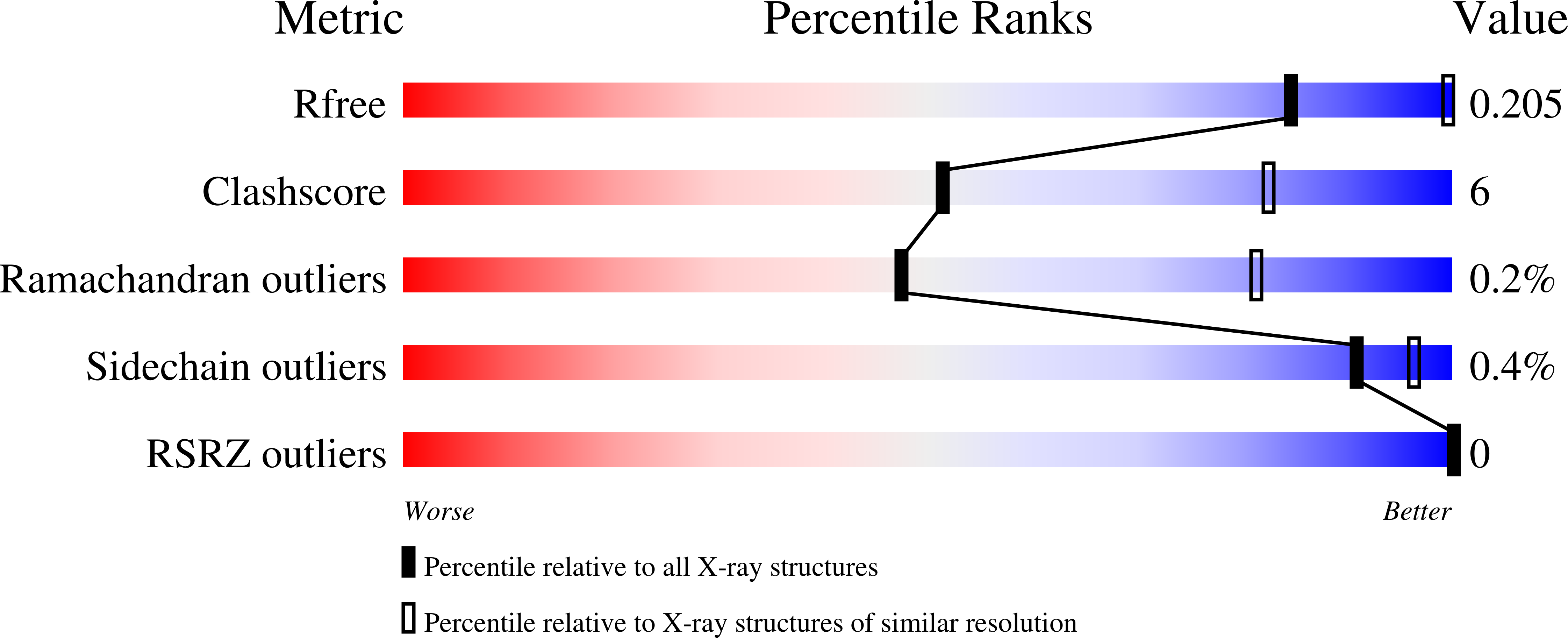
  • R-Value Free: 0.205 
  • R-Value Work: 0.183 
  • R-Value Observed: 0.184 

Starting Models: experimental
View more details

wwPDB Validation   3D Report Full Report


Ligand Structure Quality Assessment 


This is version 1.4 of the entry. See complete history


Literature

Alternative conformations of a major antigenic site on RSV F.

Jones, H.G.Battles, M.B.Lin, C.C.Bianchi, S.Corti, D.McLellan, J.S.

(2019) PLoS Pathog 15: e1007944-e1007944

  • DOI: https://doi.org/10.1371/journal.ppat.1007944
  • Primary Citation of Related Structures:  
    6DC3, 6DC4, 6DC5

  • PubMed Abstract: 

    The respiratory syncytial virus (RSV) fusion (F) glycoprotein is a major target of neutralizing antibodies arising from natural infection, and antibodies that specifically bind to the prefusion conformation of RSV F generally demonstrate the greatest neutralization potency. Prefusion-stabilized RSV F variants have been engineered as vaccine antigens, but crystal structures of these variants have revealed conformational differences in a key antigenic site located at the apex of the trimer, referred to as antigenic site Ø. Currently, it is unclear if flexibility in this region is an inherent property of prefusion RSV F or if it is related to inadequate stabilization of site Ø in the engineered variants. Therefore, we set out to investigate the conformational flexibility of antigenic site Ø, as well as the ability of the human immune system to recognize alternative conformations of this site, by determining crystal structures of prefusion RSV F bound to neutralizing human-derived antibodies AM22 and RSD5. Both antibodies bound with high affinity and were specific for the prefusion conformation of RSV F. Crystal structures of the complexes revealed that the antibodies recognized distinct conformations of antigenic site Ø, each diverging at a conserved proline residue located in the middle of an α-helix. These data suggest that antigenic site Ø exists as an ensemble of conformations, with individual antibodies recognizing discrete states. Collectively, these results have implications for the refolding of pneumovirus and paramyxovirus fusion proteins and should inform development of prefusion-stabilized RSV F vaccine candidates.


  • Organizational Affiliation

    Department of Biochemistry and Cell Biology, Geisel School of Medicine at Dartmouth, Hanover, New Hampshire, United States of America.


Macromolecules
Find similar proteins by:  (by identity cutoff)  |  3D Structure
Entity ID: 1
MoleculeChains Sequence LengthOrganismDetailsImage
Fab RSD5-Germline Heavy ChainA [auth H]230Homo sapiensMutation(s): 0 
UniProt
Find proteins for Q6N089 (Homo sapiens)
Explore Q6N089 
Go to UniProtKB:  Q6N089
Entity Groups  
Sequence Clusters30% Identity50% Identity70% Identity90% Identity95% Identity100% Identity
UniProt GroupQ6N089
Sequence Annotations
Expand
  • Reference Sequence
Find similar proteins by:  (by identity cutoff)  |  3D Structure
Entity ID: 2
MoleculeChains Sequence LengthOrganismDetailsImage
Fab RSD5-Germline Light ChainB [auth L]216Homo sapiensMutation(s): 0 
UniProt
Find proteins for Q8N355 (Homo sapiens)
Explore Q8N355 
Go to UniProtKB:  Q8N355
Entity Groups  
Sequence Clusters30% Identity50% Identity70% Identity90% Identity95% Identity100% Identity
UniProt GroupQ8N355
Sequence Annotations
Expand
  • Reference Sequence
Find similar proteins by:  (by identity cutoff)  |  3D Structure
Entity ID: 3
MoleculeChains Sequence LengthOrganismDetailsImage
RSV fusion glycoproteinC [auth F]568Human respiratory syncytial virus A2Human immunodeficiency virus 1unidentified
This entity is chimeric
Mutation(s): 7 
UniProt
Find proteins for M1E1E4 (Human immunodeficiency virus 1)
Explore M1E1E4 
Go to UniProtKB:  M1E1E4
Find proteins for P03420 (Human respiratory syncytial virus A (strain A2))
Explore P03420 
Go to UniProtKB:  P03420
Entity Groups  
Sequence Clusters30% Identity50% Identity70% Identity90% Identity95% Identity100% Identity
UniProt GroupsM1E1E4P03420
Glycosylation
Glycosylation Sites: 2
Sequence Annotations
Expand
  • Reference Sequence
Small Molecules
Ligands 2 Unique
IDChains Name / Formula / InChI Key2D Diagram3D Interactions
NAG
Query on NAG

Download Ideal Coordinates CCD File 
L [auth F],
M [auth F]
2-acetamido-2-deoxy-beta-D-glucopyranose
C8 H15 N O6
OVRNDRQMDRJTHS-FMDGEEDCSA-N
SO4
Query on SO4

Download Ideal Coordinates CCD File 
D [auth H]
E [auth L]
F [auth L]
G [auth L]
H [auth L]
D [auth H],
E [auth L],
F [auth L],
G [auth L],
H [auth L],
I [auth L],
J [auth L],
K [auth L],
N [auth F],
O [auth F],
P [auth F],
Q [auth F],
R [auth F]
SULFATE ION
O4 S
QAOWNCQODCNURD-UHFFFAOYSA-L
Experimental Data & Validation

Experimental Data

  • Method: X-RAY DIFFRACTION
  • Resolution: 3.50 Å
  • R-Value Free: 0.205 
  • R-Value Work: 0.183 
  • R-Value Observed: 0.184 
  • Space Group: I 2 3
Unit Cell:
Length ( Å )Angle ( ˚ )
a = 270.32α = 90
b = 270.32β = 90
c = 270.32γ = 90
Software Package:
Software NamePurpose
PHENIXrefinement
MOSFLMdata reduction
Aimlessdata scaling
PHASERphasing

Structure Validation

View Full Validation Report


Ligand Structure Quality Assessment 


Entry History & Funding Information

Deposition Data


Funding OrganizationLocationGrant Number
National Institutes of Health/National Institute of General Medical Sciences (NIH/NIGMS)United StatesP20GM113132

Revision History  (Full details and data files)

  • Version 1.0: 2019-07-10
    Type: Initial release
  • Version 1.1: 2020-01-01
    Changes: Author supporting evidence
  • Version 1.2: 2020-01-22
    Changes: Database references
  • Version 1.3: 2020-07-29
    Type: Remediation
    Reason: Carbohydrate remediation
    Changes: Data collection, Derived calculations, Structure summary
  • Version 1.4: 2023-10-04
    Changes: Data collection, Database references, Refinement description, Structure summary