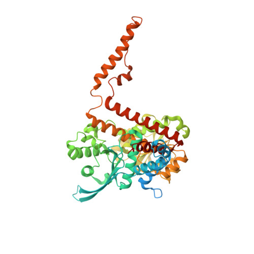
6DHM

Bovine glutamate dehydrogenase complexed with zinc


Experimental Data Snapshot

  • Method: X-RAY DIFFRACTION
  • Resolution: 3.00 Å
  • R-Value Free: 0.270 
  • R-Value Work: 0.197 
  • R-Value Observed: 0.201 

wwPDB Validation   3D Report Full Report


Ligand Structure Quality Assessment 


This is version 1.1 of the entry. See complete history


Literature

A novel mechanism of V-type zinc inhibition of glutamate dehydrogenase results from disruption of subunit interactions necessary for efficient catalysis.

Bailey, J.Powell, L.Sinanan, L.Neal, J.Li, M.Smith, T.Bell, E.

(2011) FEBS J 278: 3140-3151

  • DOI: https://doi.org/10.1111/j.1742-4658.2011.08240.x
  • Primary Citation of Related Structures:  
    6DHM, 6DHN

  • PubMed Abstract: 

    Bovine glutamate dehydrogenase is potently inhibited by zinc and the major impact is on V(max) suggesting a V-type effect on catalysis or product release. Zinc inhibition decreases as glutamate concentrations decrease suggesting a role for subunit interactions. With the monocarboxylic amino acid norvaline, which gives no evidence of subunit interactions, zinc does not inhibit. Zinc significantly decreases the size of the pre-steady state burst in the reaction but does not affect NADPH binding in the enzyme-NADPH-glutamate complex that governs the steady state turnover, again suggesting that zinc disrupts subunit interactions required for catalytic competence. While differential scanning calorimetry suggests zinc binds and induces a slightly conformationally more rigid state of the protein, limited proteolysis indicates that regions in the vicinity of the antennae regions and the trimer-trimer interface become more flexible. The structures of glutamate dehydrogenase bound with zinc and europium show that zinc binds between the three dimers of subunits in the hexamer, a region shown to bind novel inhibitors that block catalytic turnover, which is consistent with the above findings. In contrast, europium binds to the base of the antenna region and appears to abrogate the inhibitory effect of zinc. Structures of various states of the enzyme have shown that both regions are heavily involved in the conformational changes associated with catalytic turnover. These results suggest that the V-type inhibition produced with glutamate as the substrate results from disruption of subunit interactions necessary for efficient catalysis rather than by a direct effect on the active site conformation.


  • Organizational Affiliation

    Gustavus Adolphus College, St Peter, MN, USA.


Macromolecules
Find similar proteins by:  (by identity cutoff)  |  3D Structure
Entity ID: 1
MoleculeChains Sequence LengthOrganismDetailsImage
Glutamate dehydrogenase 1, mitochondrial
A, B, C, D, E
A, B, C, D, E, F
582Bos taurusMutation(s): 0 
EC: 1.4.1.2 (PDB Primary Data), 1.4.1.3 (UniProt)
UniProt
Find proteins for P00366 (Bos taurus)
Explore P00366 
Go to UniProtKB:  P00366
Entity Groups  
Sequence Clusters30% Identity50% Identity70% Identity90% Identity95% Identity100% Identity
UniProt GroupP00366
Sequence Annotations
Expand
  • Reference Sequence
Small Molecules
Ligands 4 Unique
IDChains Name / Formula / InChI Key2D Diagram3D Interactions
NDP
Query on NDP

Download Ideal Coordinates CCD File 
BA [auth E]
H [auth A]
HA [auth F]
M [auth B]
R [auth C]
BA [auth E],
H [auth A],
HA [auth F],
M [auth B],
R [auth C],
W [auth D]
NADPH DIHYDRO-NICOTINAMIDE-ADENINE-DINUCLEOTIDE PHOSPHATE
C21 H30 N7 O17 P3
ACFIXJIJDZMPPO-NNYOXOHSSA-N
GTP
Query on GTP

Download Ideal Coordinates CCD File 
CA [auth E]
I [auth A]
IA [auth F]
N [auth B]
S [auth C]
CA [auth E],
I [auth A],
IA [auth F],
N [auth B],
S [auth C],
X [auth D]
GUANOSINE-5'-TRIPHOSPHATE
C10 H16 N5 O14 P3
XKMLYUALXHKNFT-UUOKFMHZSA-N
GLU
Query on GLU

Download Ideal Coordinates CCD File 
AA [auth E]
G [auth A]
GA [auth F]
L [auth B]
Q [auth C]
AA [auth E],
G [auth A],
GA [auth F],
L [auth B],
Q [auth C],
V [auth D]
GLUTAMIC ACID
C5 H9 N O4
WHUUTDBJXJRKMK-VKHMYHEASA-N
ZN
Query on ZN

Download Ideal Coordinates CCD File 
DA [auth E]
EA [auth E]
FA [auth F]
J [auth A]
JA [auth F]
DA [auth E],
EA [auth E],
FA [auth F],
J [auth A],
JA [auth F],
K [auth A],
O [auth B],
P [auth B],
T [auth C],
U [auth C],
Y [auth D],
Z [auth D]
ZINC ION
Zn
PTFCDOFLOPIGGS-UHFFFAOYSA-N
Experimental Data & Validation

Experimental Data

  • Method: X-RAY DIFFRACTION
  • Resolution: 3.00 Å
  • R-Value Free: 0.270 
  • R-Value Work: 0.197 
  • R-Value Observed: 0.201 
  • Space Group: P 1 21 1
Unit Cell:
Length ( Å )Angle ( ˚ )
a = 121.11α = 90
b = 98.76β = 101.55
c = 165.64γ = 90
Software Package:
Software NamePurpose
PHENIXrefinement
PROTEUM PLUSdata reduction
PROTEUM PLUSdata scaling
PHENIXphasing

Structure Validation

View Full Validation Report


Ligand Structure Quality Assessment 


Entry History 

Deposition Data

  • Released Date: 2018-07-25 
  • Deposition Author(s): Smith, T.J.
  • This entry supersedes: 3MVQ

Revision History  (Full details and data files)

  • Version 1.0: 2018-07-25
    Type: Initial release
  • Version 1.1: 2024-03-13
    Changes: Data collection, Database references, Derived calculations