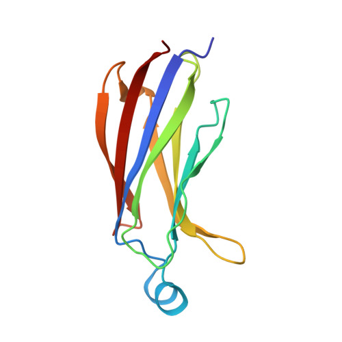
6IEJ

The C2 domain of cytosolic phospholipase A2 alpha bound to phosphatidylcholine


Experimental Data Snapshot

  • Method: X-RAY DIFFRACTION
  • Resolution: 2.21 Å
  • R-Value Free: 0.249 
  • R-Value Work: 0.224 
  • R-Value Observed: 0.226 

Starting Model: experimental
View more details

wwPDB Validation   3D Report Full Report


Ligand Structure Quality Assessment 


This is version 1.2 of the entry. See complete history


Literature

Structural basis of phosphatidylcholine recognition by the C2-domain of cytosolic phospholipase A2alpha.

Hirano, Y.Gao, Y.G.Stephenson, D.J.Vu, N.T.Malinina, L.Simanshu, D.K.Chalfant, C.E.Patel, D.J.Brown, R.E.

(2019) Elife 8

  • DOI: https://doi.org/10.7554/eLife.44760
  • Primary Citation of Related Structures:  
    6IEJ

  • PubMed Abstract: 

    Ca 2+ -stimulated translocation of cytosolic phospholipase A 2 α (cPLA 2 α) to the Golgi induces arachidonic acid production, the rate-limiting step in pro-inflammatory eicosanoid synthesis. Structural insights into the cPLA 2 α preference for phosphatidylcholine (PC)-enriched membranes have remained elusive. Here, we report the structure of the cPLA 2 α C2-domain (at 2.2 Å resolution), which contains bound 1,2-dihexanoyl- sn -glycero-3-phosphocholine (DHPC) and Ca 2+ ions. Two Ca 2+ are complexed at previously reported locations in the lipid-free C2-domain. One of these Ca 2+ ions, along with a third Ca 2+ , bridges the C2-domain to the DHPC phosphate group, which also interacts with Asn65. Tyr96 plays a key role in lipid headgroup recognition via cation-π interaction with the PC trimethylammonium group. Mutagenesis analyses confirm that Tyr96 and Asn65 function in PC binding selectivity by the C2-domain and in the regulation of cPLA 2 α activity. The DHPC-binding mode of the cPLA 2 α C2-domain, which differs from phosphatidylserine or phosphatidylinositol 4,5-bisphosphate binding by other C2-domains, expands and deepens knowledge of the lipid-binding mechanisms mediated by C2-domains.


  • Organizational Affiliation

    Structural Biology Program, Memorial Sloan-Kettering Cancer Center, New York, United States.


Macromolecules
Find similar proteins by:  (by identity cutoff)  |  3D Structure
Entity ID: 1
MoleculeChains Sequence LengthOrganismDetailsImage
Cytosolic phospholipase A2
A, B, C
127Gallus gallusMutation(s): 0 
Gene Names: PLA2G4ACPLA2PLA2G4
EC: 3.1.1.4 (PDB Primary Data), 3.1.1.5 (UniProt)
UniProt
Find proteins for P49147 (Gallus gallus)
Explore P49147 
Go to UniProtKB:  P49147
Entity Groups  
Sequence Clusters30% Identity50% Identity70% Identity90% Identity95% Identity100% Identity
UniProt GroupP49147
Sequence Annotations
Expand
  • Reference Sequence
Small Molecules
Ligands 3 Unique
IDChains Name / Formula / InChI Key2D Diagram3D Interactions
HXG
Query on HXG

Download Ideal Coordinates CCD File 
H [auth A],
L [auth B],
Q [auth C]
1,2-dihexanoyl-sn-glycero-3-phosphocholine
C20 H41 N O8 P
DVZARZBAWHITHR-GOSISDBHSA-O
CA
Query on CA

Download Ideal Coordinates CCD File 
D [auth A]
E [auth A]
F [auth A]
I [auth B]
J [auth B]
D [auth A],
E [auth A],
F [auth A],
I [auth B],
J [auth B],
K [auth B],
M [auth C],
N [auth C],
O [auth C]
CALCIUM ION
Ca
BHPQYMZQTOCNFJ-UHFFFAOYSA-N
MG
Query on MG

Download Ideal Coordinates CCD File 
G [auth A],
P [auth C]
MAGNESIUM ION
Mg
JLVVSXFLKOJNIY-UHFFFAOYSA-N
Experimental Data & Validation

Experimental Data

  • Method: X-RAY DIFFRACTION
  • Resolution: 2.21 Å
  • R-Value Free: 0.249 
  • R-Value Work: 0.224 
  • R-Value Observed: 0.226 
  • Space Group: C 2 2 2
Unit Cell:
Length ( Å )Angle ( ˚ )
a = 108.294α = 90
b = 187.388β = 90
c = 68.754γ = 90
Software Package:
Software NamePurpose
PHENIXrefinement
HKL-2000data reduction
HKL-2000data scaling
PHASERphasing

Structure Validation

View Full Validation Report


Ligand Structure Quality Assessment 


Entry History & Funding Information

Deposition Data


Funding OrganizationLocationGrant Number
National Institutes of Health/National Heart, Lung, and Blood Institute (NIH/NHLBI)United StatesHL125353

Revision History  (Full details and data files)

  • Version 1.0: 2019-05-22
    Type: Initial release
  • Version 1.1: 2022-03-23
    Changes: Author supporting evidence, Database references, Derived calculations
  • Version 1.2: 2023-11-22
    Changes: Data collection, Refinement description