6JR4

Crystal structure of a NIR-emitting DNA-stabilized Ag16 nanocluster


Experimental Data Snapshot

  • Method: X-RAY DIFFRACTION
  • Resolution: 1.80 Å
  • R-Value Free: 0.146 
  • R-Value Work: 0.126 
  • R-Value Observed: 0.128 

wwPDB Validation   3D Report Full Report


This is version 1.3 of the entry. See complete history


Literature

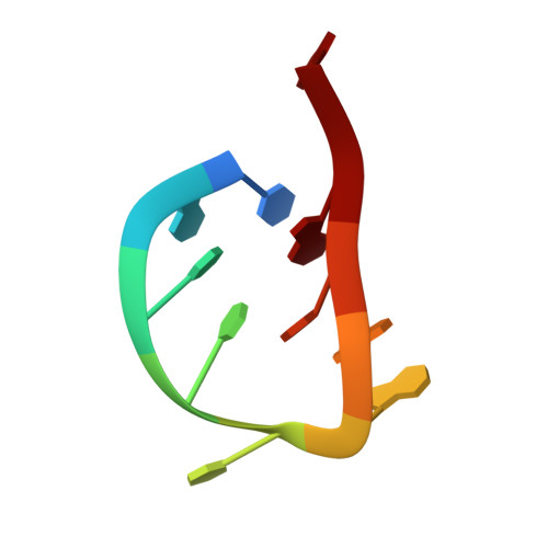
Crystal structure of a NIR-Emitting DNA-Stabilized Ag16Nanocluster.

Cerretani, C.Kanazawa, H.Vosch, T.Kondo, J.

(2019) Angew Chem Int Ed Engl 58: 17153-17157

  • DOI: https://doi.org/10.1002/anie.201906766
  • Primary Citation of Related Structures:  
    6JR4

  • PubMed Abstract: 

    DNA has been used as a scaffold to stabilize small, atomically monodisperse silver nanoclusters, which have attracted attention due to their intriguing photophysical properties. Herein, we describe the X-ray crystal structure of a DNA-encapsulated, near-infrared emitting Ag 16 nanocluster (DNA-Ag 16 NC). The asymmetric unit of the crystal contains two DNA-Ag 16 NCs and the crystal packing between the DNA-Ag 16 NCs is promoted by several interactions, such as two silver-mediated base pairs between 3'-terminal adenines, two phosphate-Ca 2+ -phosphate interactions, and π-stacking between two neighboring thymines. Each Ag 16 NC is confined by two DNA decamers that take on a horse-shoe-like conformation and is almost fully shielded from the solvent environment. This structural insight will aid in the determination of the structure/photophysical property relationship for this class of emitters and opens up new research opportunities in fluorescence imaging and sensing using noble-metal clusters.


  • Organizational Affiliation

    Department of Chemistry and NanoScience Center, University of Copenhagen, Universitetsparken 5, 2100, Copenhagen, Denmark.


Macromolecules

Find similar nucleic acids by:  Sequence   |   3D Structure  

Entity ID: 1
MoleculeChains LengthOrganismImage
DNA (5'-D(*CP*AP*CP*CP*TP*AP*GP*CP*GP*A)-3')
A, B, C, D
10synthetic construct
Sequence Annotations
Expand
  • Reference Sequence
Small Molecules
Ligands 2 Unique
IDChains Name / Formula / InChI Key2D Diagram3D Interactions
AG (Subject of Investigation/LOI)
Query on AG

Download Ideal Coordinates CCD File 
AA [auth C]
BA [auth C]
CA [auth C]
DA [auth C]
E [auth A]
AA [auth C],
BA [auth C],
CA [auth C],
DA [auth C],
E [auth A],
EA [auth C],
F [auth A],
FA [auth C],
G [auth A],
GA [auth C],
H [auth A],
HA [auth C],
I [auth A],
IA [auth C],
J [auth A],
JA [auth C],
K [auth A],
KA [auth C],
L [auth A],
LA [auth D],
M [auth A],
MA [auth D],
N [auth A],
NA [auth D],
O [auth A],
OA [auth D],
P [auth A],
PA [auth D],
Q [auth A],
QA [auth D],
R [auth B],
S [auth B],
T [auth B],
U [auth B],
V [auth B],
W [auth B],
Y [auth C],
Z [auth C]
SILVER ION
Ag
FOIXSVOLVBLSDH-UHFFFAOYSA-N
CA
Query on CA

Download Ideal Coordinates CCD File 
RA [auth D],
X [auth B]
CALCIUM ION
Ca
BHPQYMZQTOCNFJ-UHFFFAOYSA-N
Experimental Data & Validation

Experimental Data

  • Method: X-RAY DIFFRACTION
  • Resolution: 1.80 Å
  • R-Value Free: 0.146 
  • R-Value Work: 0.126 
  • R-Value Observed: 0.128 
  • Space Group: P 1 21 1
Unit Cell:
Length ( Å )Angle ( ˚ )
a = 24.241α = 90
b = 33.849β = 97.81
c = 58.863γ = 90
Software Package:
Software NamePurpose
XSCALEdata scaling
PHENIXrefinement
PDB_EXTRACTdata extraction
XDSdata reduction
PHENIXphasing

Structure Validation

View Full Validation Report


Entry History & Funding Information

Deposition Data


Funding OrganizationLocationGrant Number
Ministry of Education, Culture, Sports, Science and Technology (Japan)Japan17H03033

Revision History  (Full details and data files)

  • Version 1.0: 2019-08-28
    Type: Initial release
  • Version 1.1: 2019-11-27
    Changes: Database references
  • Version 1.2: 2020-03-25
    Changes: Data collection
  • Version 1.3: 2024-03-27
    Changes: Data collection, Database references, Derived calculations