6OIC

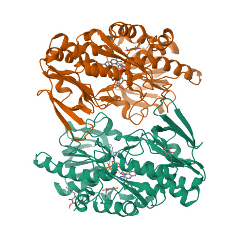
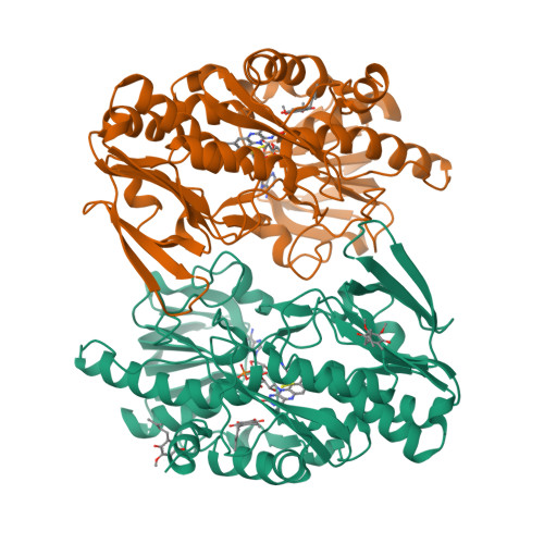
Crystal structure of human Sulfide Quinone Oxidoreductase in complex with coenzyme Q (sulfite soaked)

  • Classification: OXIDOREDUCTASE
  • Organism(s): Homo sapiens
  • Expression System: Escherichia coli
  • Mutation(s): No 

  • Deposited: 2019-04-09 Released: 2020-01-15 
  • Deposition Author(s): Banerjee, R., Cho, U.S., Kim, H., Moon, S.
  • Funding Organization(s): National Institutes of Health/National Institute of General Medical Sciences (NIH/NIGMS), National Institutes of Health/National Institute of Diabetes and Digestive and Kidney Disease (NIH/NIDDK)

Experimental Data Snapshot

  • Method: X-RAY DIFFRACTION
  • Resolution: 2.21 Å
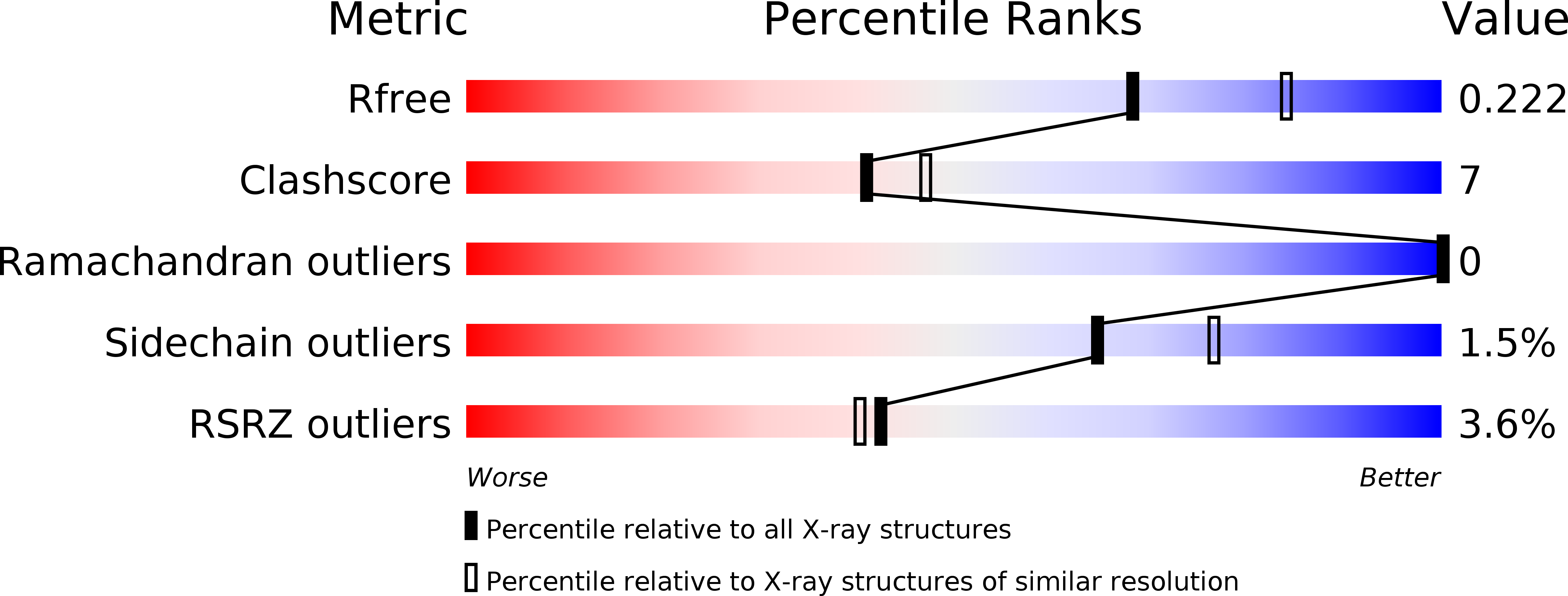
  • R-Value Free: 0.220 
  • R-Value Work: 0.182 
  • R-Value Observed: 0.184 

wwPDB Validation   3D Report Full Report


Ligand Structure Quality Assessment 

Created with Raphaël 2.3.0Worse 01 BetterLigand structure goodness of fit to experimental dataBest fitted FADClick on this verticalbar to view detailsBest fitted UQ1Click on this verticalbar to view details

This is version 1.0 of the entry. See complete history


Literature

A Catalytic Trisulfide in Human Sulfide Quinone Oxidoreductase Catalyzes Coenzyme A Persulfide Synthesis and Inhibits Butyrate Oxidation.

Landry, A.P.Moon, S.Kim, H.Yadav, P.K.Guha, A.Cho, U.S.Banerjee, R.

(2019) Cell Chem Biol 26: 1515-1525.e4

  • DOI: https://doi.org/10.1016/j.chembiol.2019.09.010
  • Primary Citation of Related Structures:  
    6OI5, 6OI6, 6OIB, 6OIC

  • PubMed Abstract: 

    Mitochondrial sulfide quinone oxidoreductase (SQR) catalyzes the oxidation of H 2 S to glutathione persulfide with concomitant reduction of CoQ 10 . We report herein that the promiscuous activity of human SQR supported the conversion of CoA to CoA-SSH (CoA-persulfide), a potent inhibitor of butyryl-CoA dehydrogenase, and revealed a molecular link between sulfide and butyrate metabolism, which are known to interact. Three different CoQ 1 -bound crystal structures furnished insights into how diverse substrates access human SQR, and provided snapshots of the reaction coordinate. Unexpectedly, the active site cysteines in SQR are configured in a bridging trisulfide at the start and end of the catalytic cycle, and the presence of sulfane sulfur was confirmed biochemically. Importantly, our study leads to a mechanistic proposal for human SQR in which sulfide addition to the trisulfide cofactor eliminates 201 Cys-SSH, forming an intense charge-transfer complex with flavin adenine dinucleotide, and 379 Cys-SSH, which transfers sulfur to an external acceptor.


  • Organizational Affiliation

    Department of Biological Chemistry, University of Michigan Medical School, Ann Arbor, MI 48109, USA.


Macromolecules
Find similar proteins by:  (by identity cutoff)  |  3D Structure
Entity ID: 1
MoleculeChains Sequence LengthOrganismDetailsImage
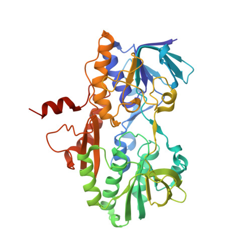
Sulfide:quinone oxidoreductase, mitochondrial
A, B
418Homo sapiensMutation(s): 0 
Gene Names: SQORSQRDLCGI-44
EC: 1.8.5 (PDB Primary Data), 1.8.5.8 (UniProt)
UniProt & NIH Common Fund Data Resources
Find proteins for Q9Y6N5 (Homo sapiens)
Explore Q9Y6N5 
Go to UniProtKB:  Q9Y6N5
PHAROS:  Q9Y6N5
GTEx:  ENSG00000137767 
Entity Groups  
Sequence Clusters30% Identity50% Identity70% Identity90% Identity95% Identity100% Identity
UniProt GroupQ9Y6N5
Sequence Annotations
Expand
  • Reference Sequence
Small Molecules
Ligands 3 Unique
IDChains Name / Formula / InChI Key2D Diagram3D Interactions
FAD
Query on FAD

Download Ideal Coordinates CCD File 
C [auth A],
H [auth B]
FLAVIN-ADENINE DINUCLEOTIDE
C27 H33 N9 O15 P2
VWWQXMAJTJZDQX-UYBVJOGSSA-N
UQ1
Query on UQ1

Download Ideal Coordinates CCD File 
D [auth A],
E [auth A],
F [auth A],
I [auth B]
UBIQUINONE-1
C14 H18 O4
SOECUQMRSRVZQQ-UHFFFAOYSA-N
H2S
Query on H2S

Download Ideal Coordinates CCD File 
G [auth A],
J [auth B]
HYDROSULFURIC ACID
H2 S
RWSOTUBLDIXVET-UHFFFAOYSA-N
Experimental Data & Validation

Experimental Data

  • Method: X-RAY DIFFRACTION
  • Resolution: 2.21 Å
  • R-Value Free: 0.220 
  • R-Value Work: 0.182 
  • R-Value Observed: 0.184 
  • Space Group: P 21 21 21
Unit Cell:
Length ( Å )Angle ( ˚ )
a = 76.101α = 90
b = 108.753β = 90
c = 131.058γ = 90
Software Package:
Software NamePurpose
PHENIXrefinement
xia2data reduction
xia2data scaling
PHASERphasing

Structure Validation

View Full Validation Report


Ligand Structure Quality Assessment 

Created with Raphaël 2.3.0Worse 01 BetterLigand structure goodness of fit to experimental dataBest fitted FADClick on this verticalbar to view detailsBest fitted UQ1Click on this verticalbar to view details

Entry History & Funding Information

Deposition Data


Funding OrganizationLocationGrant Number
National Institutes of Health/National Institute of General Medical Sciences (NIH/NIGMS)United StatesGM130183
National Institutes of Health/National Institute of Diabetes and Digestive and Kidney Disease (NIH/NIDDK)United StatesDK111465

Revision History  (Full details and data files)

  • Version 1.0: 2020-01-15
    Type: Initial release