6SXV

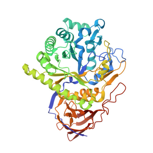
GH51 a-l-arabinofuranosidase soaked with aziridine inhibitor


Experimental Data Snapshot

  • Method: X-RAY DIFFRACTION
  • Resolution: 1.40 Å
  • R-Value Free: 0.178 
  • R-Value Work: 0.170 

Starting Model: experimental
View more details

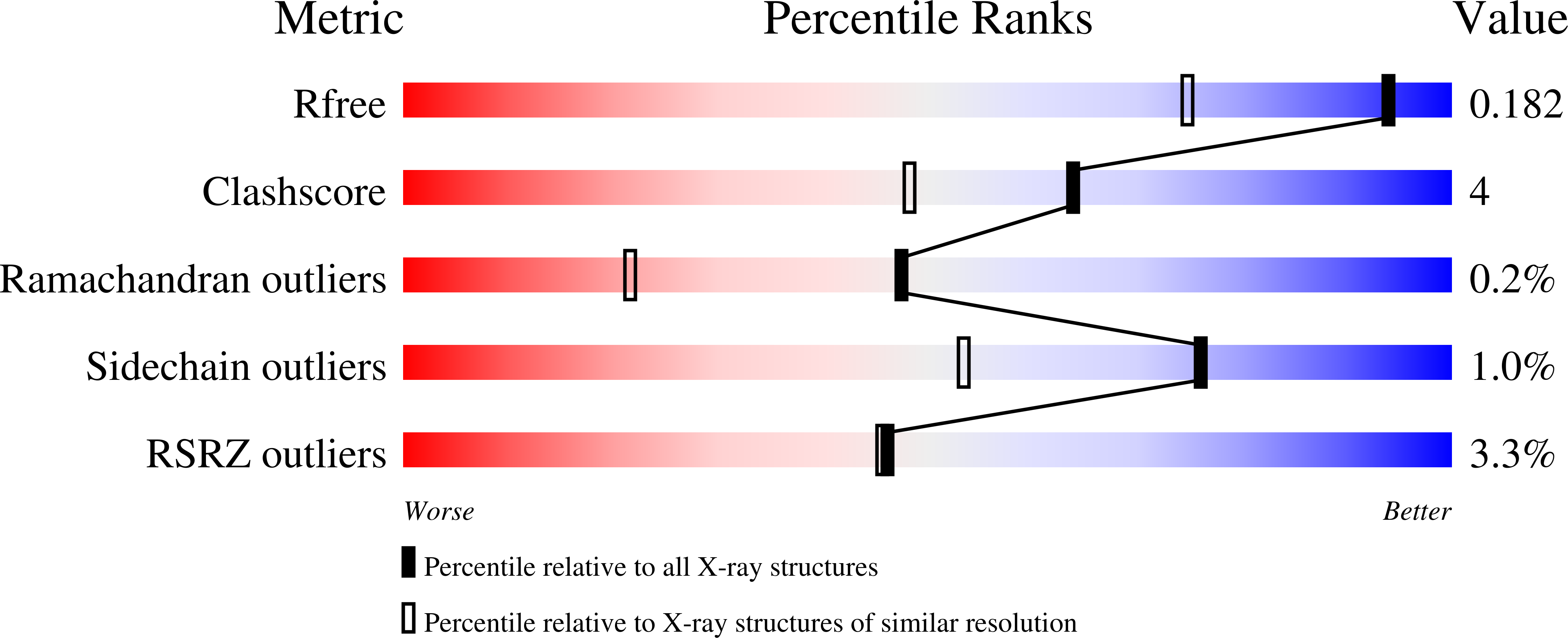
wwPDB Validation   3D Report Full Report


Ligand Structure Quality Assessment 


This is version 1.3 of the entry. See complete history


Literature

Rational Design of Mechanism-Based Inhibitors and Activity-Based Probes for the Identification of Retaining alpha-l-Arabinofuranosidases.

McGregor, N.G.S.Artola, M.Nin-Hill, A.Linzel, D.Haon, M.Reijngoud, J.Ram, A.Rosso, M.N.van der Marel, G.A.Codee, J.D.C.van Wezel, G.P.Berrin, J.G.Rovira, C.Overkleeft, H.S.Davies, G.J.

(2020) J Am Chem Soc 142: 4648-4662

  • DOI: https://doi.org/10.1021/jacs.9b11351
  • Primary Citation of Related Structures:  
    6SXR, 6SXS, 6SXT, 6SXU, 6SXV

  • PubMed Abstract: 

    Identifying and characterizing the enzymes responsible for an observed activity within a complex eukaryotic catabolic system remains one of the most significant challenges in the study of biomass-degrading systems. The debranching of both complex hemicellulosic and pectinaceous polysaccharides requires the production of α-l-arabinofuranosidases among a wide variety of coexpressed carbohydrate-active enzymes. To selectively detect and identify α-l-arabinofuranosidases produced by fungi grown on complex biomass, potential covalent inhibitors and probes which mimic α-l-arabinofuranosides were sought. The conformational free energy landscapes of free α-l-arabinofuranose and several rationally designed covalent α-l-arabinofuranosidase inhibitors were analyzed. A synthetic route to these inhibitors was subsequently developed based on a key Wittig-Still rearrangement. Through a combination of kinetic measurements, intact mass spectrometry, and structural experiments, the designed inhibitors were shown to efficiently label the catalytic nucleophiles of retaining GH51 and GH54 α-l-arabinofuranosidases. Activity-based probes elaborated from an inhibitor with an aziridine warhead were applied to the identification and characterization of α-l-arabinofuranosidases within the secretome of A. niger grown on arabinan. This method was extended to the detection and identification of α-l-arabinofuranosidases produced by eight biomass-degrading basidiomycete fungi grown on complex biomass. The broad applicability of the cyclophellitol-derived activity-based probes and inhibitors presented here make them a valuable new tool in the characterization of complex eukaryotic carbohydrate-degrading systems and in the high-throughput discovery of α-l-arabinofuranosidases.


  • Organizational Affiliation

    York Structural Biology Laboratory, Department of Chemistry, The University of York, Heslington, York YO10 5DD, U.K.


Macromolecules
Find similar proteins by:  (by identity cutoff)  |  3D Structure
Entity ID: 1
MoleculeChains Sequence LengthOrganismDetailsImage
GH51 a-l-arabinofuranosidase
A, B
502Geobacillus stearothermophilusMutation(s): 0 
Gene Names: AbfA
EC: 3.2.1.55
UniProt
Find proteins for Q9XBQ3 (Geobacillus stearothermophilus)
Explore Q9XBQ3 
Go to UniProtKB:  Q9XBQ3
Entity Groups  
Sequence Clusters30% Identity50% Identity70% Identity90% Identity95% Identity100% Identity
UniProt GroupQ9XBQ3
Sequence Annotations
Expand
  • Reference Sequence
Small Molecules
Ligands 5 Unique
IDChains Name / Formula / InChI Key2D Diagram3D Interactions
ETE
Query on ETE

Download Ideal Coordinates CCD File 
O [auth B]2-{2-[2-2-(METHOXY-ETHOXY)-ETHOXY]-ETHOXY}-ETHANOL
C9 H20 O5
ZNYRFEPBTVGZDN-UHFFFAOYSA-N
PGE
Query on PGE

Download Ideal Coordinates CCD File 
F [auth A]TRIETHYLENE GLYCOL
C6 H14 O4
ZIBGPFATKBEMQZ-UHFFFAOYSA-N
LXE (Subject of Investigation/LOI)
Query on LXE

Download Ideal Coordinates CCD File 
C [auth A],
K [auth B]
[(1~{S},2~{S},3~{S},4~{S})-2-(hydroxymethyl)-3,4-bis(oxidanyl)cyclopentyl]azanium
C6 H14 N O3
XJKPQBOZNVQXOP-VANKVMQKSA-O
PEG
Query on PEG

Download Ideal Coordinates CCD File 
E [auth A],
M [auth B]
DI(HYDROXYETHYL)ETHER
C4 H10 O3
MTHSVFCYNBDYFN-UHFFFAOYSA-N
GOL
Query on GOL

Download Ideal Coordinates CCD File 
D [auth A]
G [auth A]
H [auth A]
I [auth A]
J [auth A]
D [auth A],
G [auth A],
H [auth A],
I [auth A],
J [auth A],
L [auth B],
N [auth B],
P [auth B],
Q [auth B]
GLYCEROL
C3 H8 O3
PEDCQBHIVMGVHV-UHFFFAOYSA-N
Experimental Data & Validation

Experimental Data

  • Method: X-RAY DIFFRACTION
  • Resolution: 1.40 Å
  • R-Value Free: 0.178 
  • R-Value Work: 0.170 
  • Space Group: H 3
Unit Cell:
Length ( Å )Angle ( ˚ )
a = 178.994α = 90
b = 178.994β = 90
c = 100.893γ = 120
Software Package:
Software NamePurpose
REFMACrefinement
XDSdata reduction
Aimlessdata scaling
PHASERphasing

Structure Validation

View Full Validation Report


Ligand Structure Quality Assessment 


Entry History & Funding Information

Deposition Data


Funding OrganizationLocationGrant Number
Biotechnology and Biological Sciences Research CouncilUnited KingdomBB/R001162/1

Revision History  (Full details and data files)

  • Version 1.0: 2020-02-26
    Type: Initial release
  • Version 1.1: 2020-03-18
    Changes: Database references
  • Version 1.2: 2024-01-24
    Changes: Data collection, Database references, Derived calculations, Refinement description
  • Version 1.3: 2024-10-16
    Changes: Structure summary